Download
1 / 30
370 likes | 1.2k Vues
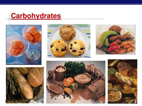
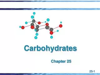
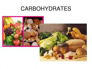
Harini Chandra. Carbohydrates. Carbohydrates, one of the four major classes of biomolecules, are aldehyde or ketone compounds with multiple hydroxyl groups. They function as energy stores, metabolic intermediates and important fuels for the body. Master Layout (Part 1). 1.

E N D
Harini Chandra Carbohydrates Carbohydrates, one of the four major classes of biomolecules, are aldehyde or ketone compounds with multiple hydroxyl groups. They function as energy stores, metabolic intermediates and important fuels for the body.
Master Layout (Part 1) 1 This animation consists of 3 parts: Part 1 – Monosaccharides Part 2 – Disaccharides & Polysaccharides Part 3 – Glycoconjugates Aldotriose Asymmetic centre Ketotriose 2 D-glyceraldehye L-glyceraldehye Dihydroxyacetone 3 Enantiomers Ketohexose Aldohexose Aldopentose 4 D-ribose D-glucose D-fructose 5 Source: Biochemistry by Lubert Stryer, 5th & 6th edition (ebook)
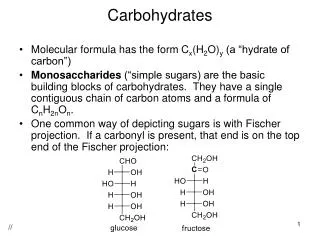
Definitions of the components:Part 1 – Monosaccharides 1 1. Monosaccharide: Monosaccharides comprise the simplest group of carbohydrates with the empirical formula (C-H2O)n. They can be either aldehydes or ketones having two or more hydroxyl groups. These monosaccharides serve as important fuel molecules and as the basic building unit for nucleic acids. 2. Aldotriose: The smallest monosaccharide having an aldehyde group and a total of 3 carbon atoms is referred to as an aldotriose. Glyceraldehyde is the simplest aldotriose. 3. Ketotriose: The smallest monosaccharide that has a ketone group with a total of 3 carbon atoms is referred to as a ketotriose. Dihydroxyacetone is the simplest ketotriose. 4. Asymmetric centre: A carbon atom that has four different groups attached to it in a tetrahedral arrangement is said to be asymmetric or chiral and gives rise to the phenomenon of optical isomerism. All monosaccharides have multiple asymmetric carbon atoms, giving rise to 2n isomers for each monosaccharide, where n refers to the number of asymmetric centres. 5. Enantiomers: Molecules with a chiral centre have a non-superimposable mirror image and the two forms of this molecule are known as enantiomers. They are designated as D and L or R (rectus) and S (sinister) depending on the arrangement of groups around the asymmetric carbon atom. R and S nomenclature is based on priority of atomic numbers of atoms directly attached to the central asymmetric centre. 2 3 4 5
Definitions of the components:Part 1 – Monosaccharides 1 6. Aldopentose: A monosaccharide having an aldehyde functional group with a total of five carbon atoms is referred to as an aldopentose. D-ribose, which is an important component of all nucleic acids is one such aldopentose. 7. Aldohexose: A monosaccharide having an aldehyde functional group with a total of six carbon atoms is referred to as an aldohexose. D-glucose is one of the most common aldohexoses. 8. Ketohexose: A sugar having a ketone functional group with a total of six carbon atoms is referred to as a ketohexose. D-fructose is the most abundant ketohexose. Ketoses have fewer asymmetric centres compared to aldoses. 9. Epimers: Sugars that differ in configuration from each other at just one asymmetric carbon atom, are referred to as epimers. Glucose and mannose are epimers at C-2 while glucose and galactose are epimers at C-4. 10. Anomers: The aldehyde or ketone functional groups can react with an alcohol to form a hemiacetal or ketal. This intramolecular reaction occurs in sugars, thereby allowing them to form cyclic structures. These are commonly represented by means of the Haworth’s projections. Upon cyclization, the aldehyde or ketone carbon becomes C1 and is referred to as the anomeric carbon atom. The configuration of groups about the anomeric carbon can result in either the alpha or beta structures, which are referred to as anomers. 2 3 4 5
Part 1, Step 1: 1 Monosaccharides – optical properties Mirror Asymmetric carbon atom 2 L-glyceraldehyde D-glyceraldehyde 3 Enantiomers 4 Action Description of the action Audio Narration All monosaccharide sugars are optically active due to the presence of asymmetric carbon atoms. The simplest aldotriose, D-glyceraldehyde, has one asymmetric centre giving rise to the D and L enantiomers, which are mirror images of each other. A compound will have 2n isomers, where n is the number of asymmetric centres.. As shown in animation. (Please redraw all figures.) First show the ball and stick figure on the left labelled as ‘D-glyceraldehyde’. Next show the appearance of a silver surface which is the ‘mirror’ followed by the reflection of the molecule as shown on the right. 5 Source: Biochemistry by Lubert Stryer, 6th edition (ebook)
Part 1, Step 2: 1 Monosaccharides – aldoses D-Erythrose D-Threose D-Glyceraldehyde 2 D-Ribose D-Xylose D-Arabinose D-Lyxose 3 D-Allose D-Altrose D-Glucose D-Gulose D-Mannose D-Iodose 4 D-Talose D-Galactose Action Description of the action Audio Narration Simple D-aldose sugars can have anywhere from three to seven carbon atoms with an aldehyde functional group. All the D-sugars have the same absolute configuration as that of D-glyceraldehyde at their asymmetric centre that is farthest away from the carbonyl carbon. The most commonly observed sugars include D-glucose, D-mannose and D-galactose. As shown in animation. (Please redraw all figures.) First show the structure on the top labelled ‘D-glyceraldehyde’ followed by the arrows on either side and appearance of next 2 structures. Next, the 2 arrows below each structure must appear sequentially along with each structure below. This must continue as shown in animation until all the structures have been displayed. 5 Source: Biochemistry by Lubert Stryer, 6th edition (ebook)
Part 1, Step 3: 1 Monosaccharides – Epimers Differ in configuration at one chiral centre - Epimers at C-2. 1 1 1 2 2 2 2 3 3 3 4 4 4 3 5 5 5 6 6 6 D-Mannose D-Glucose D-Galactose Differ in configuration at one chiral centre - Epimers at C-4. 4 Action Audio Narration Description of the action As shown in animation (Please redraw all figures.) First show the structure in the centre ‘D-glucose’ followed by the structure on the left, ‘D-mannose’. The purple rectangular boxes must then appear, along with the text box above. Next, the structure on the right must appear followed by the green rectangular boxes and the text box at the bottom. Those sugars that differ in configuration from each other about only one asymmetric carbon atom are referred to as epimers. D-Glucose and D-mannose are epimers at the second carbon atom while D-glucose and D-galactose are epimers at the fourth carbon atom. D-mannose and D-galactose, however are only diastereomers since they differ in configuration at two asymmetric centres. 5 Source: Biochemistry by Lubert Stryer, 6th edition (ebook)
Part 1, Step 4: 1 Monosaccharides - ketoses D-Sorbose 2 D-Xylulose D-Tagatose D-Erythrulose D-Fructose Dihydroxyacetone 3 D-Psicose D-Ribulose 4 Action Description of the action Audio Narration As shown in animation. (Please redraw all figures.) First the left-most figure must be shown labelled ‘dihydroxyacetone’ followed by the right arrow and the next figure ‘D-erythrulose’. Next, the two arrows must appear with each figure shown in front of the arrow. This must continue as shown in animation until all figures are displayed. Ketoses can be three, four, five or six carbon sugars with a ketone functional group. Dihydroxyacetone, the simplest ketose having 3 carbon atoms does not possess an asymmetric centre. The D configuration is therefore based on the absolute configuration of D-erythrulose, the four carbon ketose and is designated based on the asymmetric centre farthest away from the ketone group. 5 Source: Biochemistry by Lubert Stryer, 6th edition (ebook)
Part 1, Step 5: 1 ANOMERS Monosaccharides – Haworth’s projection formula Anomeric carbon H2O 2 b-D-Glucopyranose a-D-Glucopyranose D-Glucose (open chain form) 3 Chair form is more stable due to less steric hindrance in the axial positions. Boat form Chair form 4 Action Description of the action Audio Narration As shown in animation. (Please redraw all figures.) First show the figure on top left followed by appearance of green highlighting boxes and then the arrow. The structure must bend to take up the shape shown in the next figure with the highlighted groups interacting with each other. Next the reaction arrow must appear and the structures on the right must be shown. Two arrows must then appear from the figrst structure ‘b-D-glucopyranose’ and the figures below must be shown. The groups shown must be highlighted with a dotted line appearing between them in the first figure and a dotted line with a red cross mark appearing in the second followed by the callout with text. The aldehyde or ketone group of monosaccharides react with the alcohol groups to form intramolecular hemiacetals or ketals. This gives rise to stable five or six-membered rings known as furanose and pyranose respectively. The functional group carbon atom is designated as C-1 and is known as the anomeric carbon. The configuration of the groups about the anomeric carbon gives rise to the alpha and beta configurations. The chair form of the six membered pyranose ring is more stable due to minimal steric hindrance at the axial positions since they are occupied by small hydrogen atoms. 5 Source: Biochemistry by Lubert Stryer, 6th edition (ebook)
Part 1, Step 6: 1 Monosaccharides – Haworth’s projection formula ANOMERS H2O 2 b-D-Fructofuranose a-D-Fructofuranose 3 D-Fructose Open chain form 4 Action Description of the action Audio Narration As shown in animation. (Please redraw all figures.) First show the structure on the left with its label followed by the green highlighting boxes and the curved arrow. The structure must then curve so as to appear as shown in the second with the arrow mark between the highlighted regions as shown. This is followed by appearance of the arrow and the structures on the right with their labels. Although glucose is more stable in the six membered pyranose configuration, stability of fructose is greater as a five-membered furanose ring. 5 Source: Biochemistry by Lubert Stryer, 6th edition (ebook)
Part 1, Step 7: 1 Monosaccharides – Reducing nature 2 Fehling’s solution (Cu2+) 3 Glucose solution D-glucose D-gluconic acid Brown ppt. 4 Action Description of the action Audio Narration (Please redraw all figures.) First show the tube with blue solution on the left with its label. The inset must appear as being zoomed into and the first structure must be shown. Then the hand with the micropipette must be shown and drops must fall from this into the tube and dissolve on falling. Once a few drops fall, a brown mass (precipitate) must appear at the bottom of the tube. When this happens, the arrow in the reaction must appear followed by the figure on the right. As shown in animation. A simple test for identifying sugars such as glucose is by the Fehling’s test. The free aldehyde group provides the sugars with a reducing nature thereby bringing about reduction of a solution of cupric ions. This results in an easily identifiable brown precipitate. 5 Source: Biochemistry by Lubert Stryer, 6th edition (ebook)
Part 1, Step 8: 1 Modified monosaccharides N-glycosidic bond O-glycosidic bond 2 Methyl-a-D-glucopyranoside b-L-fucose (Fuc) 3 Sialic acid (Sia) (N-Acetylneuraminate) b-D-Acetylgalactosamine (GalNAc) b-D-Acetylglucosamine (GlcNAc) 4 Action Description of the action Audio Narration As shown in animation. (Please redraw all figures.) Show each structure appearing sequentially with the regions highlighted as shown and their respective labels. The reactive anomeric carbon atom can be modified by reaction with alcohols or amines to form adducts. Reaction of glucose with methanol gives the corresponding methyl glucopyranoside with the formation of an O-glycosidic bond. When the anomeric carbon is linked to an amine via its nitrogen atom, it results in formation of an N-glycosidic bond. Several other modified monosaccharides such as fucose, N-acetyl glucosamine etc are present which serve various structural and functional roles. 5 Source: Biochemistry by Lubert Stryer, 6th edition (ebook)
Master Layout (Part 2) 1 This animation consists of 3 parts: Part 1 – Monosaccharides Part 2 – Disaccharides & Polysaccharides Part 3 – Glycoconjugates Sucrose 2 Glycosidic bond 3 Starch & glycogen 4 Cellulose 5 Source: Biochemistry by Lubert Stryer, 6th edition (ebook)
Definitions of the components:Part 2 – Disaccharides & Polysaccharides 1 1. Disaccharide: Two monosaccharide units joined together by means of an O-glycosidic linkage forms a disaccharide. Three of the most common disaccharides include maltose, sucrose and lactose. 2. Polysaccharide: Several monosaccharide units joined together by glycosidic bonds form a polysaccharide. These help in maintaining structural integrity and serve as fuel reserves in organisms. Some of the most common polysaccharides include starch, the nutritional reservoir in plants, glycogen – the fuel storage form in animal cells and cellulose, which is the most important structural elements in plants. 3. Glycosidic bond: The bond formed by interaction between the hydroxyl group of one monosaccharide with the hydroxyl group, aldehyde or ketone group of another monosaccharide, with the subsequent elimination of water is known as the glycosidic bond. 4. Hemiacetal: The interaction between an aldehyde or ketone group with a hydroxyl group with the elimination of water results in the formation of a hemiacetal or ketal. In sugars, intramolecular hemiacetals and ketals are formed by cyclization of the sugar molecules. 5. Homopolymer: When all the monosaccharide units of a polysaccharide are the same, it is said to be a homopolymer. The most common homopolymers are starch and glycogen. 6. Heteropolymer: When the repeating monosaccharide units of the polysaccharide are different, they are said to be heteropolymers. 2 3 4 5
Definitions of the components:Part 2 – Disaccharides & Polysaccharides 1 7. Starch: The nutritional reserve of plants is starch, which is composed of two components – amylose and amylopectin. Amylose consists of linear, unbranched chains of D-glucose residues linked by a-1,4 glycosidic linkages. Amylopectin, however, is a branched polymer with an a-1,6 glycosidic linkage present around every 30 residues. Starch is rapidly degraded by the enzyme amylase. 8. Cellulose: Cellulose plays a major structural role in plants and consists of linear chains of glucose residues linked together by b-1,4 glycosidic bonds. These chains formed by b-linkages have very high tensile strength and can be digested by the enzyme cellulase, which is not inherent in mammals. 9. Glycogen: Glycogen is the storage form of glucose in animal cells. It is structurally similar to starch, with glucose residues being joined by a-1,4 glycosidic linkages. Glycogen has more extensive branching than starch with branch points being observed around every 10 residues. These allow the molecules to be stored in a very compact way in the cells. 10. Chitin: Chitin is another structural polysaccharide that is a homopolymer of N-acetyl-D-glucosamine residues joined together by b-1,4 glycosidic linkages. It is commonly found on the exoskeleton of insects. 2 3 4 5
Part 2, Step 1: 1 Disaccharides - formation Alcohol Hemiacetal 2 a-D-glucose b-D-glucose H2O H2O 3 Glycosidic bond Maltose a-D-Glucopyranosyl-(14)-a-D-glucopyranose 4 Action Description of the action Audio Narration Disaccharides are formed by the condensation reaction between two monosaccharide units. The release of a molecule of water results in the formation of a glycosidic bond between the two residues. Shown in this example is the formation of maltose, a disaccharide that is composed of two units of glucose linked by an a-14 glycosidic bond. Maltose is hydrolyzed into its individual units by the enzyme maltase. (Please redraw all figures.) First show the structures on top left & top right with the ‘+’ sign in between. These two structures must move close to each other and the red highlighted regions must be removed as ‘H2O’ and the resulting structure at the bottom shown with its labels. As shown in animation. 5 Source: Biochemistry by Lubert Stryer, 6th edition (ebook)
Part 2, Step 2: 1 Common disaccharides 2 Sucrose a-D-glucopyranosyl-(12)-b-D-fructofuranose 3 Lactose b-D-Galactopyranosyl-(14)-a-D-glucopyranose 4 Action Description of the action Audio Narration As shown in animation. (Please redraw all figures.) Show the sequential appearance of the figures above as shown. Table sugar or sucrose is a disaccharide composed of one unit of glucose and one of fructose linked by a b-12 bond. It is a non-reducing sugar since the aldehyde group of glucose and ketone group of fructose are involved in formation of the glycosidic linkage. It can be cleaved by the enzyme sucrase. Lactose, the sugar component of milk, is made up of one unit of galactose and one of glucose linked by an a-14 linkage. It can be cleaved by the enzyme lactase, also known as b-galactosidase. 5 Source: Biochemistry by Lubert Stryer, 6th edition (ebook)
Part 2, Step 3: 1 Polysaccharides: Starch, a homopolymer of D-glucose residues Reducing end Non-reducing end a-1, 4-glycosidic linkage Starch granules 2 Amylose 3 Branch a-(16) branch point Main chain Amylopectin 4 Action Description of the action Audio Narration Starch granules, which form the major nutritional reserve of plants, are composed of two components – amylose and amylopectin. Amylose consists of linear, unbranched chains of D-glucose residues linked by a-1,4 glycosidic linkages. Amylopectin, however, is a branched polymer with an a-1,6 glycosidic linkage present around every 30 residues. Starch is rapidly degraded by the enzyme amylase. As shown in animation. (Please redraw all figures.) First show the figure on the left with the label for ‘starch granules’. This region must then be zoomed into and the structures on the right must be shown. 5 Source: Biochemistry by Lubert Stryer, 6th edition (ebook)
Part 2, Step 4: 1 Glycogen is a highly branched molecule with branch points every 10 residues. Polysaccharides: Glycogen, a homopolysaccharide of D-glucose residues a-1, 4-glycosidic linkage Glycogen granules 2 3 a-(16) branch point 4 Action Description of the action Audio Narration Glycogen is the storage form of glucose in animal cells. It is structurally similar to start with glucose residues being joined by a-1,4 glycosidic linkages. Glycogen has more extensive branching than starch with branch points being observed around every 10 residues. These allow the molecules to be stored in a very compact way in the cells. As shown in animation. (Please redraw all figures.) First show the figure on the left with the label ‘glycogen granules’. This must then be zoomed into and the structure on the right must be shown. This is followed by appearance of the brown text star on the top. 5 Source: Biochemistry by Lubert Stryer, 6th edition (ebook)
Part 2, Step 5: 1 Polysaccharides: Cellulose & Chitin 2 N-acetyl-D-glucosamine residues b-1, 4-glycosidic linkage Cellulose 3 Chitin b-(14) linkage 4 Action Description of the action Audio Narration As shown in animation. (Please redraw all figures.) Show the sequential appearance of the two structures with their labels as depicted. Cellulose plays a major structural role in plants and consists of linear chains of glucose residues linked together by b-1,4 glycosidic bonds. These chains formed by b-linkages have very high tensile strength and can be digested by the enzyme cellulase, which is not inherent in mammals.Chitin is another structural polysaccharide that is a homopolymer of N-acetyl-D-glucosamine residues joined together by b-1,4 glycosidic linkages. It is commonly found on the exoskeleton of insects. 5 Source: Biochemistry by Lubert Stryer, 6th edition (ebook)
Master Layout (Part 3) 1 This animation consists of 3 parts: Part 1 – Monosaccharides Part 2 – Disaccharides & Polysaccharides Part 3 – Glycoconjugates Carboxyl terminus Proteoglycan structure Gly Glycolipid X 2 Gly Sugar residues (b 13) (b 13) (b 14) (b 14) (b 13) Glycosidic bonds in glycoproteins (GlcA GalNAc 4S)n GlcA Gal Gal Xyl Ser 3 Chondroitin sulphate Asn Ser Core protein Amino terminus Lipid 4 O-linked GalNAc N-linked GlcNAc 5 Source: Biochemistry by A.L.Lehninger, 4th edition (ebook); Biochemistry by Lubert Stryer, 6th edition (ebook)
Definitions of the components:Part 3 – Glycoconjugates 1 1. Glycosaminoglycan: Glycosaminoglycans are heteropolysaccharide repeating units found on animal cell surfaces and in the extracellular matrix. They are composed of disaccharide units that contain a derivative of an amino sugar and at least one of the sugars in the unit has a negatively charged carboxylate or sulphate group. 2. Proteoglycan: These are structural elements that are composed of glycosaminoglyan units linked to proteins. The proteoglycan, aggrecan, is a major component of cartilage along with the protein, collagen, where it serves as a shock absorber. Degradation of aggrecan and collagen can lead to osteoarthritis. 3. Glycoprotein: Carbohydrate groups are often covalently attached to proteins to form glycoproteins. The sugar residues are typically attached to the amide nitrogen atom of the aspargine side chain or to the oxygen atom of the serine or threonine side chain. These glycoproteins are components of cell membranes and have a variety of functions in cell adhesion processes. 4. Glycolipid: Carbohydrate moieties can also be covalently linked with various lipids. Glycolipids are membrane components bearing a hydrophilic head group and a hydrophobic lipid tail. 5. Glycosyl transferase: These are a specific class of enzymes that are responsible for the transfer or sugar residues onto other substrates. They transfer the sugar in its activated state such as UDP-glucose to other molecules such as other monosaccharides, polysaccharides or amino acid side chains of proteins. 2 3 4 5
Part 3, Step 1: 1 2 Chondroitin-6-sulphate Keratan sulphate Glycosaminoglycans 3 Heparin Hyaluronate Dermatan sulphate 4 Action Description of the action Audio Narration As shown in animation. (Please redraw all figures.) First show the heading in the centre followed by sequential appearance of all the structures with their labels as depicted in animation. Glycosaminoglycans are heteropolysaccharide components of extracellular matrix spaces along with other fibrous proteins. They are linear polymers that are composed of disaccharide repeating units of which one residue is always a derivative of an amino sugar such as N-acetyl glucosamine or N-acetyl galactosamine. They also contain a negatively charged sulphate or carboxylate group.Common glycosaminoglycans include chondrotin sulphate, hyaluronate, heparin etc. 5 Source: Biochemistry by Lubert Stryer, 6th edition (ebook)
Part 3, Step 2: 1 Glycosyltransferase reaction 2 Substrate to be glycosylated UDP-glucose Glycosyl transferase 3 Glycosylated substrate UDP 4 Action Description of the action Audio Narration As shown in animation. (Please redraw all figures.) First show the structures on top with their labels. Next show the arrow mark with the label. The pink structure must then be broken from the remaining black structure at the position indicated by the dotted line and the blue ‘X’ must be attached there in place of the ‘O’ as shown in the figure below. And the blue ‘H’ must be attached to the red ‘O’ linked to remaining black structure. Glycosyl transferases are specific enzymes that bring about transfer of activated sugar residues onto other substrates. The activated sugar linked to a nucleotide moiety such as UDP is cleaved and covalently linked with the substrate which could either be another monosaccharide unit, a polysaccharide or the serine or aspargine side chains of proteins. 5 Source: Biochemistry by Lubert Stryer, 6th edition (ebook)
Part 3, Step 3: 1 Cartilage is made of the proteoglycan, aggrecan, which serves as a shock absorber. Proteoglycan structure Carboxyl terminus Gly X 2 Gly (b 13) (b 13) (b 14) (b 14) (b 13) (GlcA GalNAc 4S)n GlcA Gal Gal Xyl Ser Protein Chondroitin sulphate Glycosaminoglycan Core protein 3 Amino terminus 4 Action Description of the action Audio Narration As shown in animation. Glycosaminoglycans can be linked to proteins to form various proteoglycans. These molecules have a variety of functions in tissue organization, development of specialized tissues and for modulation of ligand interactions with cell surface receptors. Aggrecan is a proteoglycan aggregate consisting of many core proteins bound to a single hyaluronate molecules; they serve as shock absorbers in cartilage. (Please redraw all figures.) First show the colored rectangular structure with all the labels. Next show the appearance of the vertical chain on the side as depicted in animation followed by appearance of the speech bubble with text. 5 Source: Biochemistry by A.L.Lehninger, 4th edition (ebook)
Part 3, Step 4: 1 Glycoproteins Blood groups Ser Asn Fuc Fuc Gal GalNAc Fuc 2 Gal Gal Gal GlcNAc GlcNAc GlcNAc 3 Gal Gal Gal O-linked GalNAc N-linked GlcNAc O antigen A antigen B antigen 4 Action Description of the action Audio Narration As shown in animation. Carbohydrate groups are often covalently attached to proteins to form glycoproteins. The sugar residues are typically attached to the amide nitrogen atom of the aspargine side chain or to the oxygen atom of the serine or threonine side chain. These glycoproteins are components of cell membranes and have a variety of functions in cell adhesion processes. The ABO blood groups arise due to differing carbohydrate structures on the surface of the blood cells. (Please redraw all figures.) First show the two structures on the left appearing one after the other. Then show the figures on the right with each structure appearing sequentially as depicted in the animation. 5 Source: Biochemistry by Lubert Stryer, 6th edition (ebook)
Part 3, Step 5: 1 Glycolipids Lipopolysaccharide Sugar residues 2 3 Bacterial cell Cell membrane Lipid 4 Action Description of the action Audio Narration As shown in animation. Carbohydrate moieties can also be covalently linked with various lipids. Glycolipids are membrane components bearing a hydrophilic head group and a hydrophobic lipid tail. Lipopolysaccharides are a predominant feature on the outer membrane of gram negative bacteria, which consist of fatty acid chains bound to sugar residues. (Please redraw all figures.) First show the ‘bacterial cell’ on the left. The region marked in red must be zoomed into and the next structure must be shown. The red circle must then appear with its label on this structure, which must be further zoomed into to show the rightmost structure. 5 Source: Biochemistry by A.L.Lehninger, 4th edition (ebook)
Interactivity option 1:Step No: 1 1 A common chemical test to detect the presence of starch is to treat it with a solution of iodine. The reaction is believed to occur due to charge transfer between iodine molecules that fit inside the amylose coils and the glucose residues of amylose. Starche that contain less amylose content do not give a prominent colour change. Drag and drop the iodine molecules into the crevices of the amylose helix shown to view the reaction. 2 I3- I3- 3 Amylose Blue colour indicating presence of starch I3- I3- I3- 4 Results Interacativity Type Options Boundary/limits The user must drag and drop the round structures into the curved spaces to view the reaction as shown in the animation. Once the user drags all the round figures into the crevices, the colour of the chain must turn blue as shown in the figure on the right. Drag & drop. (Please redraw all figures.) User must drag & drop the round structures into the curved spaces. 5
Questionnaire 1 1. Which of the following monosaccharides does not have an asymmetric centre? Answers: a) D-Glyceraldehyde b) D-Erythrulose c) Dihydroxyacetone d) D-Ribulose 2. Which of the following sugars is a C-2 epimer of glucose? Answers: a) Galactose b) Mannose c) Iodose d) Allose 3. Chitin is a homopolysaccharide of which of the following monosaccharide residues? Answers:a) N-acetyl-D-glucosamine b) a-D-glucose c) b-D-glucose d) D-fructose 4. Lactose is a disaccharide composed of which of these monosaccharide units? Answers:a) 2 units of Glucose b) Glucose & fructose c) 2 units of Galactose d) Glucose & galactose 5. The glycosaminoglycan found in aggrecan is? Answers:a) Chondroitin-6-sulphate b) Heparin c) Keratan sulphate d) Hyaluronate 2 3 4 5
Links for further reading Books: Biochemistry by Stryer et al., 6th edition Biochemistry by A.L.Lehninger et al., 4th edition Biochemistry by Voet & Voet, 3rd edition