Download
1 / 6
60 likes | 216 Vues
Supplemental Figure 1. The vast majority of alterations are shared between primary and metastatic tumors. Of the 434 total mutations, 344 (79%) were shared between patient-matched tumors (purple dots). Mutations private to primary are in pink, and mutations private to metastasis are in blue.

E N D
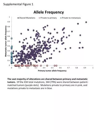
Supplemental Figure 1 The vast majority of alterations are shared between primary and metastatic tumors. Of the 434 total mutations, 344 (79%) were shared between patient-matched tumors (purple dots). Mutations private to primary are in pink, and mutations private to metastasis are in blue.
Supplemental Figure 2 A B Convergent evolution of genes was found in colon primary and metastatic tumors. (A) Patient 38 has two different TP53 mutations: p.R248Q in the primary and p.Y163* in the metastasis. (B) Patient 7 has two hotspot mutations in PIK3CA: p.E542K in the primary and p.E545K in the metastasis.
Supplemental Figure 3 Patient 19 had a metastasis-specific activating mutation in MEK1.
Supplemental Figure 4 MAP2K1 K57N MAP2K1 Q56P MAP2K1 A106T MAP2K1 Mock MAP2K1 F53L p-ERK (S202/T204) ERK The MAP2K1 p.A106T mutation is not an activating mutation. MAP2K1 p.A106T GFP-tagged plasmids were transfected into 293H cells and behave as wildtype MAP2K1. GFP* GAPDH *MEK1 plasmid tagged with GFP
Supplemental Figure 5 Months followup Survival difference based on mutational concordance. There is no survival difference between patients with concordant and discordant primary and metastatic tumors.
Supplemental Figure 6 A B D C E F H G Whole genome sequencing shows that IMPACT results predict for concordance or discordance at the larger genome level. Patient 54 (A-D) is completely concordant at the mutation (A) and copy number level (B) by IMPACT, and WGS shows a high degree of concordance (C) with all nonsense and splice site mutations in both primary and metastasis (D, red dots). Patient 19 (E-H) was discordant by IMPACT (E-F) and showed tremendous discordance at the whole genome level (G-H).