Understanding Gel Electrophoresis of DNA: Principles and Applications
Gel electrophoresis is a fundamental technique used to separate DNA fragments based on size, taking advantage of the negative charge of DNA molecules. When placed in an electric field, these molecules migrate towards the anode. This process occurs in medium gel, usually agarose or acrylamide, where smaller fragments travel faster through narrow passages. The DNA bands can be visualized using ethidium bromide and UV light post-separation. Common buffer systems include TBE and TAE, making gel electrophoresis an essential tool in molecular biology and genetics for analyzing DNA samples.

Understanding Gel Electrophoresis of DNA: Principles and Applications
E N D
Presentation Transcript
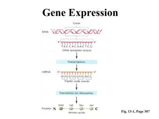
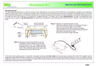
Worksheet IX.7 Genial- Agarose gel electrophoresis Gel Electrophoresis The DNA fragments produced by a restriction enzyme can be separated by size using the fact that DNA due to the backbone of phosphoric acid residues is negatively charged and moves in response to an electric field. If the terminals of an electrical power source are connected to the opposite ends of a horizontal tube containing a DNA solution, then the DNA molecules will move toward the positive end of the tube at a rate thatdepends on the electric field strength andon the shape and size of the molecules. The movement of charged molecules in an electric field is called electrophoresis. The type of electrophoresis most commonly used in genetics is gel electrophoresis. An experimental arrangement for gel electrophoresis of DNA is shown in Figures1 and 2 (Hartl and Jones 2000). A thin slab of a gel, usually agarose or acrylamide, is prepared containing small slots (wells) into which samples are placed. An electric field is applied, and the negatively charged DNA molecules penetrate and move through the gel toward the anode (the positively charged electrode). A gel is a complex molecular network that contains narrow, tortuous passages, so smaller DNA molecules pass through more easily; hence the rate of movement increases as the size of the DNA fragment decreases (Hartl and Jones 2000). Figure 1. Apparatus for gel electrophoresis (open) Liquid gel is allowed to harden in an shaped mould with a comb in it to form wells (slots) for the samples. After electrophoresis the DNA-fragments, located at various positions in the gel, are made visible by treatment with ethidium bromide and UV-light. (Hartl and Jones 2000, slightly changed) comb Figure 2. Apparatus for gel electrophoresis (closed and connected to a power supply) Commonly, gel electrophoresis runs by ~ 100 voltages and DNA fragments moves according its charge toward the anode. Often used buffer systems for agarose gel electrophoresis include TBE and TAE. page: