Endocirne glands
1.03k likes | 1.31k Vues
Endocirne glands. David Kachlík. Endocrine glands Glandulae endocrinae. one of regulation systems hormone (gr. horman – to arise) chemical messanger produced by endocrine gland and transported in blood to target organs proteins (polypeptides) – insuline amines – adrenaline

Endocirne glands
E N D
Presentation Transcript
Endocirne glands David Kachlík
EndocrineglandsGlandulaeendocrinae • one of regulation systems • hormone (gr. horman – to arise) • chemical messanger produced by endocrine gland and transported in blood to target organs • proteins (polypeptides) –insuline • amines –adrenaline • steroids –estrogenes
Endocrine glandsHistory Thomas Wharton • 1614-1673 • Adenographia • first detailed description of glands
Endocrine glandsHistory Ernest Henry Starling • 1866-1927 • general schemes of „endocrine secretion“ • used the already exsiting word „hormones“
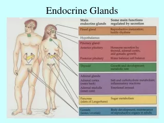
Endocrine glands arrangement • glands • disseminated cells • neuroendocrine cells
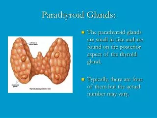
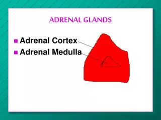
Endocrine glands – list • hypothalamus (hypothalamus) • pituitary gland (hypophysis; gl. pituitaria) • thyroid gland (glandula thyroidea) • parathyroid bodies(gll. parathyroideae) • suprarenal glands, adrenals (gll. suprarenales) • pancreatic (Langerhans‘) island (insulae pancreaticae) • pineal glands, epiphysis (gl. pinealis; corpus pineale)
Hypothalamus + hypophysis Systema hypothalamo-hypophysiale Hypothalamo-hypophysial axis
Hypophysis; Glandula pituitariaHistory • Galenos – mucus production for nasal mucosa • Schneider – 1655 refused Galenos‘ idea • Minkowski, Hutchinson – connection between growth disroders and hypohysial hypertrophy • Cushing – explained the function „a conducter of endocrine system, a prime minister“
Hypothalamus • basal part of diencephalon, basally to 3rd ventricle • function • information collection center from body and surroundings • highest autonomic center • part of limbic system • manages other endocrine glands • corpora mammillaria, tuber cinereum, infundibulum, hypophysis
Hypothalamus • anterior hypothalamus – ncl. magnocellularis • ncl. paraventricularis + supraopticus – oxytocine and vasopressin (ADH) • middle hypothalamus (tuber cinereum) – ncl. parvocelularis • ncl. arcuatus and surroundings – management of adenohypophysis • posterior hypothalamus
Hypothalamus – hormones • ncl. arcuatus – production • eminetia mediana – releasing into first capillary network • releasing hormones = liberins • SRH, PRH, GnRH, TRH, CRH • inhibiting hormones = statins • somatostatin, PIH (= dopamine)
Hypophysis – anatomy „double glands“ - two different tissues • two lobes • anterior = adenohypophysis • posterior = neurohypophysis • located within sella turcica ossis sphenoidalis • transsphenoidal operation approach • covered with dura mater – diaphragma sellae • foramen diaphragmatis Pacchioni – transmits infundibulum
Hypophysis – anatomy • anterior lobe (adenohypophysis; lobus anterior) • pars distalis (principalis) – largets part (75%) • pars intermedia – between both lobes • pars tuberalis – cranially at infudibulum • posterior lobe (neurohypohysis; lobus posterior) • lobus nervosus (pars nervosa) – proper posterior lobe • infundibulum – connection to hypothalamus
Hypohysis – blood supply hypophysial portal system • a. hypophysialis inferior (from pars cavernosa ACI to neurohypophysis) • a. hypophysialis superior (from pars cerebralis ACI via hypothalamus to adenohypophysis) • vv. hypophysiales into sinus cavernosus
Hypophysis – development • pouch of Ratke • ectoderm→ anterior lobe • 3rd week: in the roof of stomodeum • pouch towards diencephalon • separation of pouch, proliferation of anterior wall • pouch of diencephalic base • neuroectoderm → posterior lobe • differentiation into v pituicytes (glia)
Pars distalis adenohypophysis • cords of cells (chordae endocrinocytorum) • fenestrated capillaries inbetween cords • 3 types of cells in HE staining: • acidophilic • basophilic • PAS-positive • chromophobe • no granule, undifferentiated elements
Pars distalis – acidophilic cells • α – cells (endocrinocytus somatotropicus) • large granulee, GER • zone without granules around nucleus (GA) • somatotrophin (human growth hormone, GH) • ε – cells (endocrinocytus prolactinicus) • usually small, infrequent • multiplication in gravidity and lactation • little granules (larger in gravidity) • prolactin (PRL)
Pars distalis – basophilic cells • β1 – cells (endocrinocytus corticotropicus) • large granules at cytoplasmatic membrane • ACTH, β-MSH, Met-enkefalin, endorphine • β2 – cells (endocrinocytus thyrotropicus) • large cells, small granules at BM • TSH • δ – cells (endocrinocytus gonadotropicus) • large cells, middle granules • FSH, LH (lutropin)
Adenohypophysis modified Azan imunoperoxidase reaction to LH • HE
Pars tuberalis adenohypophysis • encircles the infundibulum • frequent capillaries • majority: δ-cells • few β2-cells
Pars intermedia adenohyophysis • rudimentary • cells form trabecules • basophilic cells • follicle of Ratkhe can be formed
Posterior lobe = Neurohypophysis • eminentia mediana • floor of the 3rd ventricle • frequent nonmyelinized nerve fibers • infundibulum • tractus hypothalamohypophysialis • neurofibra neurosecretoria (+ vesicula neurosecretoria) = nonmyelinized nerve fibers • some terminate at capillaries • lobus nervosus (pars nervosa)
Lobus nervosus neurohypophysis • nerve fibers • axons of hypothalamic neurons • corpuscula neurosecretoria (bodies of Herring) – accumulation of granules • oxytocin + ADH (adiuretin, antidiuretic hormone, vasopressin) • pituicytes (pituicyti) • glial cells • capillaries (synapsis neurohaemalis)
Examination and diseases • CT • hormone levels in blood • tumors of hypophysis – usually benign, hormonactive • Sheehan‘s syndrome – postpartal bleeding into hypophysis
Thyroid gland – history • Galenos – makes the pharynx wet inside hltanu • Paracelsus – goiter + cretenism • Wharton (1614-1673) – decoration of female neck • Simon (1844) – endocrine glands • Murray (1891) – application of thyroid gland extraction • Baumann (1895) – thyroid glands contains iodium compounds
Thyroid gland – anatomy • thyroxine T4, triiodothyronine T3 • calcitonine • located at level of C6-C7 • 2 lobes – lobus dexter + sinister • isthmus • on 2nd-4th tracheal cartilage • capsula fibrosa – 2 layers – stroma • parenchyma + lobuli
Thyroid gland – blood vessels • a. thyroidea superior (← a. carotis externa) • a. thyroidea inferior (← truncus thyrocervicalis) • crossing with n. laryngeus recurrens • a. thyroidea ima Neubaueri (← arcus aortae) • 2% • vv. thyroideae superiores et mediae Lichačevae-Kocheri→ vv. jugularis interna • vv. thyroideae inferiores → plexus thyroideus impar → v. brachiocephalica sinistra
Thyroid gland – development • from 24th day • pouch of primitiv pharynx endoderm • both relative and absolute descent →ductus thyroglossus • foramen caecum • gll. thyroideae accessoriae • lobes formation • lobus pyramidalis • ligamentum suspensorium gl. thyroideae / musculus levator glandulae thyroideae (smooth)
Thyroid gland – histogenesis • solid endodermal structure • ingrowth of surrounding mesenchyme and vessels • ingrowth of ultimopharyngeal (ultimobranchial) bodies • 10th week: division of cells into groups • simple epithelium around lumen • 11th week: colloid production starts
Thyroid gland – structure • capsula fibrosa • stroma • septa (between lobules) • lobus lobulus folliculus • follicles (50–900 μm) • spheric • simple epithelium of follicular cells • contains colloidum (colloid) – thyreoglobulin • follicular cell (thyrocytus T) • parafollicular cell (thyrocytus C)
Follicular cells (Thyrocytus T) • spheric nucleus • large gER (basally) and mitochondria • numerous lysosomes • thyreoglobulin, cleavage of T4 and T3
1. iodine pump using ATP transport hte iodine form blood to colloid 2. + 3. synthesis of thyreoglobulin and peroxidase, storage in one secretory vesicle and their release into the colloid by exocytosis 4. iodination of thyreoglobulin by peroxidase within the colloid and formation of iodinethyreoglobulin endocytosis of iodinethyreoglobulin 5. fusion of primary lysosoma with this vesicle proteolysis of iodinethyreoglobulin into T3, T4 and other fragments release of T3 and T4 into circulation 6. binding to transport plasmatic protein (TBP) Synthesis of thyroid gland hormones
Parafollicular cells (Thyrocytus C) • C-cells • derived from neural crest from ultimopharyngeal body • located between follicles (individually or in groups) • larger, brighter • rich gER, GA, MIT • granule – spheric, dark • prodcution and storages of calcitonine
Thyroid gland examination • utrasound • scintigraphy with radioactive iodine131
Thyroid gland – diseases • less than 10 μg of iodine daily goiter from lack of iodine • hypothyroidism • cretenism (children) – screening of newborns • myxoedema (adults) • autoimunne – thyroiditis of Hashimoto • hyperthyroidism (thyreotoxicosis) • autoimunne – exophthalmic goiter = disease of Graves-Basedow