Download
1 / 59
590 likes | 618 Vues
Uncover the interaction of proteins with mechanical forces and understand protein denaturation and mechanical unfolding at a molecular level. Learn about key proteins like fibronectin and cadherin, and the impact of forces on protein structure and function. Explore the Freely Jointed Chain (FJC) model of entropic elasticity and protein engineering techniques.

E N D
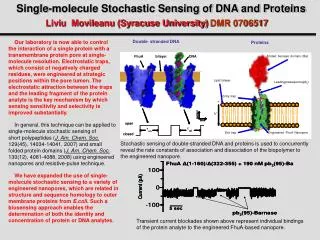
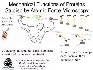
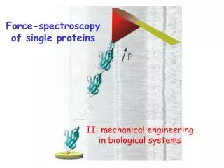
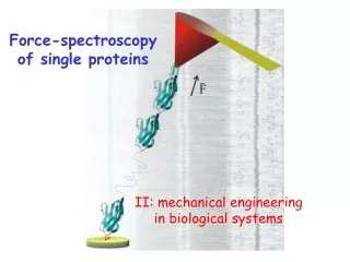
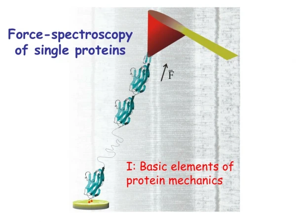
Force-spectroscopy of single proteins I: Basic elements of protein mechanics
Are proteins exposed to mechanical forces in-vivo? extracellular matrix connector complex P0 fibronectin Schwann cell Cytoskeleton: actin, etc. tenascin cell fascilin Cadherin NCAM Basal Lamina: tenascin fibronectin etc. Anchor proteins Dystrophin Cytoskeleton titin synapse cell-matrix muscle F ion channels
Mechanical forces are a natural protein denaturant. Denaturant = Force (physiological) Proteins unfold and extend under a stretching force reaction coordinate (Dx; end-to-end length) If we then relax the force, the protein folds!
We can stretch a single protein and measure how does the restoring force changes with the extension. mirrors laser Photodiode (Force) cantilever protein linear actuator (extension) .
Modified VEECO detector head Physik Instrumente “pico cube” actuator
Polypeptides are freely jointed segments with complex chemical groups branching out on the sides.
Increased entropy makes polymers look crooked and pushes them to collapse
d Path of a particle undergoing Brownian motion
A polymer looks like the path of a particle in Brownian motion l
If we had a 1 meter long polypeptide…………. 1 meter it would spontaneously collapse into .
x6 x3 x4 x7 x5 x2 x1 x F l1 l2 l3 F l6 l7 li l5 l4 The Freely Jointed Chain (FJC) model of entropic elasticity
Ten thousand water molecules at room temperature Switch to and activate VMD cluster program
Folded proteins have a bonded structure that helps them resist the constant bombardment caused by Brownian motion. Small protein ubiquitin cartoon representation
The bonds that hold the protein together can be though of as an energy barrier that has to be overcome to unravel the protein.
Igor Demonstration of analysis with models of polymer elasticity
Much of what we see can be explained with a simple model(for now)
How often does Brownian motion overcomes the energy barrier and causes the protein to unfold spontaneously ? Conformational change E kT end-to-end length t0 ~ 10-12 seconds
How long do we have to wait for Brownian motion to overcome an energy barrier ? pico seconds 1067years seconds Biology Too strong Brownian death E =
Control in Biology is accomplished by reducing energy barriers. Then, unlikely events will occur over short time periods DE
After mechanical unfoldinga protein can refold without errors (mostly)
Misfolding traps the linker between I27 modules
With all these measurements wecompile a rough energy landscape
How much does a single amino acid contributes to the contour length ?(or what is li for a protein)
1.51 Å 1.33 Å 1.46 Å Lcontour= 4.30 Å The end-to-end length of an amino acid vs its contour length Lend-to-end= 3.6 Å
Varying the location of the disulfide bonds across the I27 structure allows us to measure the contour length of a single amino acid