Download
1 / 56
560 likes | 602 Vues
Unravel the mechanical intricacies of proteins like titin and fibronectin, pivotal in cellular structure and activity. Understand the design and function of key protein modules and extracellular matrix components through advanced analysis and models. Dive into the fascinating world of protein elasticity and cell mechanics.

E N D
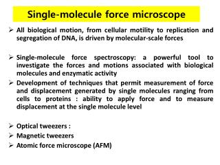
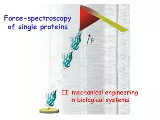
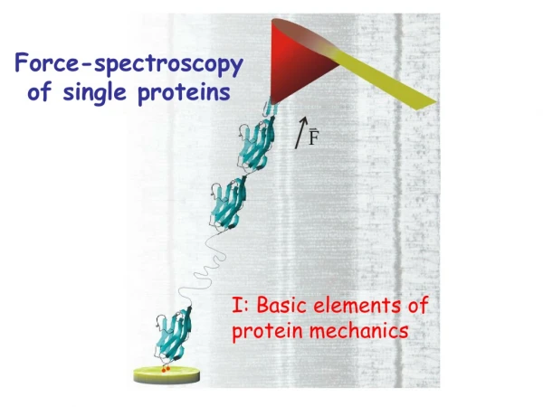
Force-spectroscopy of single proteins II: mechanical engineering in biological systems
Igor Demonstration of analysis with models of polymer elasticity
Titin: a complex mechanical protein A B C D Adapted from Linke, 2007, Cardiovascular Research (in press)
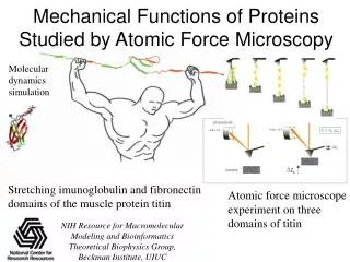
Measuring the extensibility of titin in a single isolated cardiac fiber
V11P V15P V13P wt Y9P
Understand the mechanical design of titin in humans Understand the molecular design of its modules Create titin phenotypes in mice
A complex web of proteins and polysaccharides that provides the mechanical scaffold for organs and tissues ECM cell membrane
Fibronectin: a major, cell binding component of the ECM NMR structure of 10F3. The RGD residues are identified in the picture.
Mechanical unfolding of protein domains helps to keep the cells mechanically bonded. Mechanical hierarchies define the triggers of cellular activity Cell binding cryptic binding cryptic binding
Mechanical design of the extracellular matrix:polysaccharides
Polysaccharides cellulose amylose
0.55 nm 0.45 nm If we mechanically stretch a sugar ring, it gets longer by switching from a chair to a boat conformation
Ubiquitin chains form a mechanical signallingsystem in cells