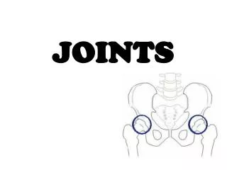
Joints
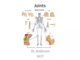
Joints. Classification of Joints. Functional classification (Focuses on amount of movement) Synarthroses (immovable joints) Amphiarthroses (slightly movable joints) Diarthroses (freely movable joints) Structural classification

Joints
E N D
Presentation Transcript
Classification of Joints • Functional classification (Focuses on amount of movement) • Synarthroses (immovable joints) • Amphiarthroses (slightly movable joints) • Diarthroses (freely movable joints) • Structural classification (Based on the material binding them and presence or absence of a joint cavity) • Bony fusion • Fibrous • Cartilagenous • Synovial
Fibrous joints • Bones connected by fibrous tissue: dense regular connective tissue • No joint cavity • Slightly immovable or not at all • Types • Sutures • Syndesmoses • Gomphoses
Sutures • Only between bones of skull • Fibrous tissue continuous with periosteum • Ossify and fuse in middle age: now technically called “synostoses”= bony junctions
Syndesmoses • In Greek: “ligament” • Bones connected by ligaments only • Amount of movement depends on length of the fibers: longer than in sutures
Gomphoses • Is a “peg-in-socket” • Only example is tooth with its socket • Ligament is a short periodontal ligament
Cartilagenous joints • Articulating bones united by cartilage • Lack a joint cavity • Not highly movable • Two types • Synchondroses (singular: synchondrosis) • Sympheses (singular: symphesis)
Synchondroses • Literally: “junction of cartilage” • Hyaline cartilage unites the bones • Immovable (synarthroses) • Examples: • Epiphyseal plates • Joint between first rib’s costal cartilage and manubrium of the sternum
Sympheses • Literally “growing together” • Fibrocartilage unites the bones • Slightly movable (amphiarthroses) • Resilient shock absorber • Provide strength and flexibility • Hyaline cartilage on articular surfaces of bones to reduce friction • Examples • Intervertebral discs • Pubic symphysis of the pelvis
Synchondroses and sympheses Also pubic symphsis
Synovial joints • Include most of the body’s joints • All are diarthroses (freely movable) • All contain fluid-filled joint cavity
General Structure of Synovial Joints • Articular cartilage • Hyaline • Spongy cushions absorb compression • Protects ends of bones from being crushed • Joint (synovial) cavity • Potential space • Small amount of synovial fluid
General structure of synovial joints (cont.) 3. Articular (or joint) capsule • Two layered • Outer*: fibrous capsule of dense irregular connective tissue continuous with periosteum • Inner*: synovial membrane of loose connective tissue (makes synovial fluid) • Lines all internal joint surfaces not covered by cartilage* * * *
General structure of synovial joints (cont.) 4. Synovial fluid • Filtrate of blood • Contains special glycoproteins • Nourishes cartilage and functions as slippery lubricant • “Weeping” lubricatioin 5. Reinforcing ligaments (some joints) • Capsular (most) – thickened parts of capsule • Extracapsular • Intracapsular
General structure of synovial joints (cont.) 6. Nerves • Detect pain • Monitor stretch (one of the ways of sensing posture and body movements) 7. Blood vessels • Rich blood supply • Extensive capillary beds in synovial membrane (produce the blood filtrate)
Some joints… • Articular disc or meniscus (literally “crescent”) • Only some joints • Those with bone ends of different shapes or fitting poorly • Some to allow two kinds of movement (e.g. jaw) • Of fibrocartilage • Examples: knee TMJ (temporomandibular joint) sternoclavicular joint
Bursae and tendon sheaths • Contain synovial fluid • Not joints but often associated with them • Act like ball bearings • Bursa means “purse” in Latin • Flattened sac lined by synovial membrane • Where ligaments, muscles, tendons, or bones overlie each other and rub together • Tendon sheath • Only on tendons subjected to friction
Joint stability • Articular surfaces • Shape usually plays only minor role • Some deep sockets or grooves do provide stability • Ligaments • Usually the more, the stronger the joint • Can stretch only 6% beyond normal length before tear • Once stretched, stay stretched • Muscle tone • Constant, low level of contractile force • Keeps tension on the ligaments • Especially important at shoulders, knees, arches of foot
Movements allowed by synovial joints • Gliding • Angular movements: hor i the angle between two bones DO TOGETHER • Flexion • Extension • Abduction • Adduction • Circumduction • Rotation • Special movements
Pronation Supination Dorsiflexion Plantar flexion Inversion Eversion Protraction Retraction Elevation Depression Opposition Special movements
Synovial joints classified by shape(of their articular surfaces) • Plane (see right) • Hinge (see right) • Pivot • Condyloid • Saddle • Ball-and-socket
Selected synovial joints Shoulder (glenohumeral) joint • Stability sacrificed for mobility • Ball and socket: head of humerus with glenoid cavity of scapula • Glenoid labrum: rim of fibrocartilage • Thin, loose capsule • Strongest ligament: coracohumeral • Muscle tendons help stability • Disorders Rotator cuff muscles add to stability Biceps tendon is intra-articular
Elbow joint • Hinge: allows only flexion and extension • Annular ligament of radius attaches to capsule • Capsule thickens into: • Radial collateral ligament • Ulnar collateral ligament • Muscles cross joint • Trauma
Wrist jointTwo major joint surfacesSeveral ligaments stabilize • Radiocarpal joint • Between radius and proximal carpals (scaphoid and lunate) • Condyloid joint • Flexion extension adduction, abduction, circumduction • Intercarpal or midcarpal joint • Between the proximal and distal rows of carpals
Hip (coxal) joint • Ball and socket • Moves in all axes but limited by ligaments and deep socket • Three ext. ligaments “screw in” head of femur when standing • Iliofemoral • Pubofemoral • Ischiofemoral
Acetabular labrum diameter smaller than head of femur • Dislocations rare • Ligament of head of femur supplies artery • Muscle tendons cross joint • Hip fractures common in elderly because of osteoporosis
Knee joint • Largest and most complex joint • Primarily a hinge • Compound and bicondyloid: femur and tibia both have 2 condyles • Femoropatellar joint shares joint cavity • At least a dozen bursae • Prepatellar • Suprapatellar
Lateral and medial menisci • “torn cartilage” • Capsule absent anteriorly • Capsular and extracapsular ligaments • Taut when knee extended to prevent hyperextension
Patellar ligament • Continuation of quad tendon • Medial and lateral retinacula • Fibular and tibial collateral ligaments • Called medial and lateral • Extracapsular • Oblique popliteal • Arcuate popliteal
Cruciate ligaments • Cross each other (cruciate means cross) • Anterior cruciate (ACL) • Anterior intercondylar area of tibia to medial side of lateral condyl of femur • Posterior cruciate • Posterior intercondylar area of tibia to lateral side of medial condyl • Restraining straps • Lock the knee
Knee injuries • Flat tibial surface predisposes to horizontal injuries • Lateral blow: multiple tears • ACL injuries • Stop and twist • Commoner in women athletes • Heal poorly • Require surgery
Ankle joint • Hinge joint • Distal tibia and fibula to talus • Dorsiflexion and plantar flexion only • Medial deltoid ligament • Lateral ligaments: 3 bands • Anterior talofibular • Posterior talofibular • Calcaneofibular • Anterior and posterior tibiofibular (syndesmosis)
Temporomandibular joint (TMJ) • Head of mandible articulates with temporal bone • Disc protects thin mandibular fossa of temporal bone • Many movements Demonstrate movements together • Disorders common
Sternoclavicular joint • Saddle joint • Only other example is trapezium and metacarpal 1 (thumb), allowing opposion • Sternum and 1st costal (rib) cartilage articulate with clavicle • Very stable: clavicle usually breaks before dislocation of joint • Only bony attachment of axial skeleton to pectoral girdle Demonstrate movements together
Disorders of joints • Injuries • Sprains • Dislocatios • Torn cartilage • Inflammatory and degenerative conditions • Bursitis • Tendinitis • Arthritis • Osteoarthritis (“DJD” – degenerative joint disease) • Rheumatoid arthritis (one of many “autoimmune” arthritites) • Gout (crystal arthropathy)