Understanding the Layers and Functions of Epidermis and Epithelial Tissues
This document provides an overview of the epidermis and the types of epithelial tissues within the integumentary system. It explains the classification of epithelial tissues based on structure, including simple and stratified types, as well as cell shapes being squamous, cuboidal, or columnar. The characteristics of epithelial tissues such as polarity, cellularity, and avascular nature are described. Additionally, key examples of simple squamous, cuboidal, and columnar epithelia are presented, along with their functions and locations, such as in the lungs, kidneys, and gall bladder.

Understanding the Layers and Functions of Epidermis and Epithelial Tissues
E N D
Presentation Transcript
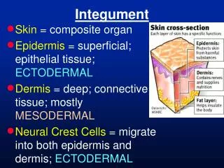
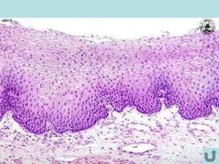
Skin (Integument) Figure 5.3 Epidermis
Layers of the Epidermis: STRATA = LAYERS C G S B D C G S B D Epidermis
Tissues • Groups of cells similar in structure and function • The four types of tissues • Epithelial • Connective • Muscle • Nerve
Epithelial Tissue • covers the body surfaces • lines the lumen of organs • lines the body cavities • forms glands and lines ducts
Epithelial Tissue - characteristics • Cellularity – • Special contacts – tight / desmosomes • Polarity – apical / basal • Supported by connective tissue – lamina • Avascular but innervated – • Regenerative –
Classification of Epithelia • Simple or stratified Figure 4.1a
apical basal
Classification of Epithelia • Squamous, cuboidal, or columnar • DESCRIPTION Figure 4.1b
Simple Squamous DESCRIPTION FUNCTIONS PRESENCE- glomeruli, alveoli, etc. Figure 4.2a
C AL Squamous Type II cell
Lumen of alveolus Simple squamous of alveolus Simple squamous of capillary Respiratory membrane Lumen of alveolus (bar = 1um)
Kidney (Glomerulus) Simple squamous epithelium (arrow) lines the glomerulus. Bar = 50um
Epithelia: Simple Cuboidal • DESCRIPTION • FUNCTIONS • PRESENCE- Figure 4.2b
RESORB FILTER SECRETE 2,000L 180L EXCRETE 1.5L
Epithelia: Simple Columnar DESCRIPTION FUNCTIONS PRESENCE Figure 4.2c
Gall BladderSimple columnar epithelium lines the gall bladder. Note the underlying connective tissue with blood vessels. Bar = 100 Microns Gall BladderSimple columnar epithelium lines the gall bladder. Note the underlying connective tissue with blood vessels. Bar = 100 Microns.
DESCRIPTION FUNCTIONS PRESENCE Figure 4.2d
Trachea The pseudostratified columnar epithelium of the trachea is ciliated and has goblet cells. Bar = 50 um
Pseudostratified columnar epithelium with cilia and goblet cells