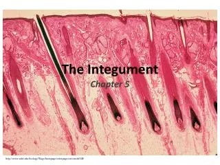
The Integument
The Integument. Chapter 5. http://www.udel.edu/biology/Wags/histopage/colorpage/cin/cinshf.GIF. Skin (Integument). Consists of three major regions Epidermis —superficial region Dermis —middle region Hypodermis (superficial fascia)—deepest region

The Integument
E N D
Presentation Transcript
The Integument Chapter 5 http://www.udel.edu/biology/Wags/histopage/colorpage/cin/cinshf.GIF
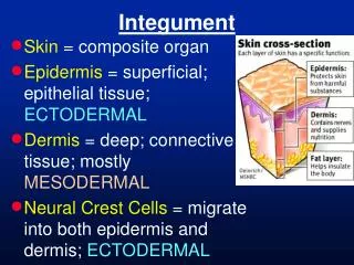
Skin (Integument) • Consists of three major regions • Epidermis—superficial region • Dermis—middle region Hypodermis (superficial fascia)—deepest region • Subcutaneous layer deep to skin (not technically part of skin) • Mostly adipose tissue
Hair shaft Dermal papillae Subpapillary vascular plexus Epidermis Papillary layer Pore Appendages of skin Dermis • Eccrine sweat gland Reticular layer • Arrector pili muscle • Sebaceous (oil) gland Hypodermis (superficial fascia) • Hair follicle Nervous structures • Hair root • Sensory nerve fiber Cutaneous vascular plexus • Pacinian corpuscle Adipose tissue • Hair follicle receptor (root hair plexus) Figure 5.1
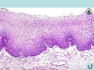
Epidermis • Keratinized stratified squamous epithelium • Cells of epidermis • Keratinocytes • Melanocytes • Epidermal dendritic (Langerhans) cells • Tactile (Merkel) cells http://www.technion.ac.il/~mdcourse/274203/slides/Skin/6-Langerhans%20Cells.jpg
Layers of the Epidermis: Stratum Basale (Basal Layer) • Deepest epidermal layer firmly attached to the dermis • Single row of stem cells • Also called stratum germinativum: cells undergo rapid division • Journey from basal layer to surface • Takes 25–45 days
Layers of the Epidermis: Stratum Spinosum (Prickly Layer) • Cells contain a weblike system of intermediate prekeratin filaments attached to desmosomes • Abundant melanin granules and dendritic cells
Layers of the Epidermis: Stratum Granulosum (Granular Layer) • Thin; three to five cell layers in which the cells flatten • Keratohyaline and lamellated granules accumulate
Layers of the Epidermis: Stratum Lucidum (Clear Layer) • Only in thick skin • Thin, transparent band superficial to the stratum granulosum • A few rows of flat, dead keratinocytes http://legacy.owensboro.kctcs.edu/gcaplan/anat/notes/161_epidermis.gif
Layers of the Epidermis: Stratum Corneum (Horny Layer) • 20–30 rows of dead, flat, keratinized membranous sacs • Three-quarters of the epidermal thickness • Functions • Protects from abrasion and penetration • Waterproofs • Barrier against biological, chemical, and physical assaults
Dermis • Strong, flexible connective tissue • Cells include fibroblasts, macrophages, and occasionally mast cells and white blood cells • Two layers: • Papillary • Reticular
Hair shaft Dermal papillae Subpapillary vascular plexus Epidermis Papillary layer Pore Appendages of skin Dermis • Eccrine sweat gland Reticular layer • Arrector pili muscle • Sebaceous (oil) gland Hypodermis (superficial fascia) • Hair follicle Nervous structures • Hair root • Sensory nerve fiber Cutaneous vascular plexus • Pacinian corpuscle Adipose tissue • Hair follicle receptor (root hair plexus) Figure 5.1
Layers of the Dermis • Papillary layer • Areolar connective tissue with collagen and elastic fibers and blood vessels • Dermal papillae • Reticular layer • ~80% of the thickness of dermis • Collagen and elastic fibers (dense irregular connective tissue)
Layers of the Dermis Epidermis Papillary Dermis Reticular
Skin Markings • Friction Ridges • Increase gripping ability of fingers and feet • Cleavage Lines • Separations b/w less dense areas of collagen fibers • Incisions made parallel to cleavage lines heal more readily
Skin Color • Three pigments contribute to skin color: • Melanin • Yellow to reddish-brown to black, responsible for dark skin colors • Carotene • Yellow to orange, most obvious in the palms and soles • Hemoglobin • Responsible for the pinkish hue of skin
Appendages of the Skin • Derivatives of the epidermis • Sweat glands • Oil glands • Hairs and hair follicles • Nails
Sweat Glands • Two main types of sweat glands • Eccrine sweat glands • abundant on palms, soles, and forehead • Apocrine sweat glands • confined to axillary and anogenital areas • Ceriminous glands – earwax • Mammary glands – secret milk
Sweat pore Eccrine gland Sebaceous gland Duct Dermal connective tissue Secretory cells (b) Photomicrograph of a sectioned eccrine gland (220x) Figure 5.5b
Sebaceous (Oil) Glands • Widely distributed • Most develop from hair follicles • Become active at puberty • Secrete Sebum
Hair • Functions • Alerting the body to presence of insects on the skin • Guarding the scalp against physical trauma, heat loss, and sunlight • Distribution • Entire surface except palms, soles, lips, nipples, and portions of external genitalia • Consists of dead keratinized cells • Hair pigments: melanins (yellow, rust brown, black)
Follicle wall • Connective tissue root sheath • Glassy membrane • External epithelial root sheath • Internal epithelial root sheath Hair shaft Hair • Cuticle • Cortex • Medulla Arrector pili (a) Diagram of a cross section of a hair within its follicle Sebaceous gland Hair root Hair bulb Figure 5.6a
Hair Follicle • Extends from the epidermal surface into dermis • Two-layered wall: outer connective tissue root sheath (dermis), inner epithelial root sheath (epidermis) • Hair bulb: expanded deep end • Hair follicle receptor (root hair plexus) • Sensory nerve endings around each hair bulb • Stimulated by bending a hair • Arrector pili • Smooth muscle attached to follicle • Responsible for “goose bumps”
Follicle wall • Connective tissue root sheath • Glassy membrane Hair shaft • External epithelial root sheath • Internal epithelial root sheath Arrector pili Hair root • Cuticle Sebaceous gland • Cortex • Medulla Hair root Hair matrix Hair bulb Hair papilla Subcutaneous adipose tissue Photomicrograph of longitudinal view of the hair bulb of the follicle (160) Figure 5.6c
Hair • Types • Vellus—pale, fine body hair of children and adult females • Terminal—coarse, long hair of eyebrows, scalp, axillary, and pubic regions (and face and neck of males) • Thinning/Baldness • Alopecia • True (frank) baldness http://thebeautybrains.com/wp-content/uploads/2007/08/baby-hair.jpg
Structure of a Nail • Scalelike modification of the epidermis on the distal, dorsal surface of fingers and toes
Functions of the Integumentary System • Protection—three types of barriers • Chemical, physical/mechanical, biological • Body temperature regulation • Cutaneous sensations • Metabolic functions • Blood reservoir • Excretion
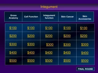
Skin Cancer • Risk factors • Overexposure to UV radiation • Frequent irritation of the skin • Three major types: • Basal cell carcinoma • Squamous cell carcinoma • Melanoma
Burns • Heat, electricity, radiation, certain chemicals • Immediate threat: • Dehydration and electrolyte imbalance, leading to renal shutdown and circulatory shock • Rule of Nines
Burns • Partial Thickness • First degree • Epidermal damage only • Second degree • Epidermal and upper dermal damage • Full Thickness • Third Degree • Entire thickness of skin
Developmental Aspects: Old Age • Epidermal replacement slows • Subcutaneous fat and elasticity decrease • Increased risk of cancer http://1.bp.blogspot.com/-f_6MgLAfZb4/T2mjLJxVr7I/AAAAAAAAAYc/DcoYDZHP8HM/s1600/old-age-bebo-dot-com.jpg