INTEGUMENT
INTEGUMENT. Surface Anatomy. Palpation Bony landmarks Dermatomes Neural assessment. Integument Histology. Epidermis: Stratified squamous epithelium Resting on: Basement membrane Resting on: Dermis: Dense irregular connective tissue. Epidermis.


INTEGUMENT
E N D
Presentation Transcript
Surface Anatomy • Palpation • Bony landmarks • Dermatomes Neural assessment
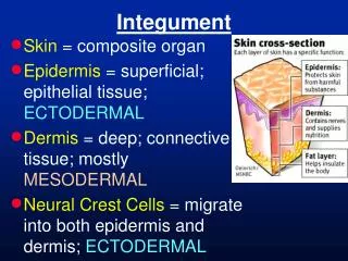
Integument Histology • Epidermis: Stratified squamous epithelium Resting on: • Basement membrane Resting on: • Dermis: Dense irregular connective tissue
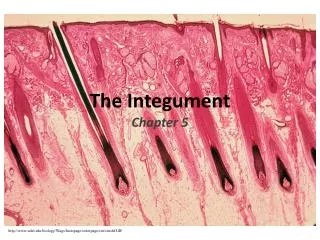
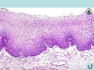
Epidermis • The epidermis is a stratified squamous epithelium. • It is made up of many layers of cells. • The stratum germinativum is the deepest layer: Area of high mitotic activity.
Epidermis • The stratum corneum is the most superficial layer: The cells in this layer are dead and keratinized. • Between the stratum germinativum and the stratum corneum are several transitional layers represented by cells from the stratum germinativum that are transforming into dead, keratinized cells.
Epidermis • The epidermis is innervated. • The epidermis is avascular.
Dermis • The dermis is the deepest region of the integument. • The dermis is classified as dense irregular connective tissue • The dermis has an abundance of collagen fibers • There may also be some elastic fibers: Decrease with age.
Dermis • The dermis is vascularized. • Refer to Figure 1 in your course packet.
Thick Skin vs. Thin Skin • Classification into thin and thick skin depends on the structure of the epidermis. • Layers of epidermis are well-formed in thick skin. • Layers of epidermis are not as well-formed or thick in thin skin.
Thick Skin • Thick skin is found only on the palms of the hands and the soles of the feet. • The epidermis of thick skin is 0.4 – 0.6 mm thick • Thick skin has no hair follicles.
Thin Skin • Thin skin is found over the rest of the body. • The epidermis of thin skin is 0.075 – 0.150 mm thick. • Total skin thickness is 0.5 – 3 mm thick.
Skin Thickness • Thickest skin found on back (= thin skin) • Thinnest skin found on eyelids (= thin skin) • Thicker on extensor surfaces than flexor surfaces.
Superficial Fascia: Synonyms • Subcutaneous fascia • Superficial fascia • Hypodermis • SubQ
Superficial Fascia • Consists of loose bundles of collagen and elastic fibers with variably sized aggregations of lipocytes (fat cells) • May be loosely or tightly attached • Supports cutaneous nerves and blood vessels
Deep Fascia • Synonyms: Membranous fascia Investing fascia • Usually several thin layers of tough collagen material • Tightly adherent to muscles, bones, tendons, etc.
Cutaneous Derivatives • Glands. • Hairs. • Nails.
Glands • Glands are epithelial structures • Glands are classified according to the presence or absence of a secretory duct: Exocrine Endocrine
Epidermal Glands • Sudoriferous glands • Sebaceous glands • Ceruminous glands • Mammary glands
Sudoriferous Glands • Are long, simple, tubular glands. • Their method of secretion is merocrine .
Sebaceous Glands • Are holocrine . • Sebaceous glands are associated with hair follicles.
Ceruminous Glands • Are located in the external auditory canal. • Secrete ear wax.
Mammary Glands • Are modified sweat glands • Method of secretion is apocrine
Hairs • Hairs develop during 3rd month of gestation. • The earliest fine embryonic hair = lanugo. • Lanugo is Shed before birth except around eyebrows, scalp, and eyelids.
Hairs • A new downy coat of hair appears a few months after birth. • This new coat is called vellus. • Vellus is converted to terminal hair at puberty: Vellus represents 95% of the hair coverage in males. Vellus represents 35% of the hair coverage in females.
Parts of a Hair • Shaft: Made up of dead cornified epidermal cells. • Follicle: Derived from both epidermis and dermis. • Dermal papilla with matrix.
Parts of a Hair • Arrector pili muscle. • Sebaceous glands. • Hair bulb and connective tissue papilla.
Hair Growth • Anlagen Active growth: Scalp hair = 2-3 years Eyebrow hair = 3-4 months
Hair Function and Location • Hair follicles are innervated, and hairs serve as sensory receptors. • Hairs are found everywhere except palms, soles, dorsal distal phalanges, anal and urogenital apertures
Nails • Ungis: Modified stratum corneum Flattened Avascular and not innervated Travels over a nail bed guided by lateral nail grooves • Matrix: Stratum germinativum produces ungis • Subungis
Melanocytes • Found in deep layers of epidermis • Derived from nervous system components • Form: Melanosomes: Passed off to keratinocytes (cells of epidermis). Phagocytized by keratinocytes.
Melanocytes • All individuals produce same number of melanosomes. • Skin color depends on number of remaining melanosomes.
Langer’s Lines • Represent tension lines created by orientation of collagen fibers in the dermis of the skin. • Used by surgeons as guides for incisions: Incisions normally made parallel to Langer’s lines
Dermatomes • Specific region of skin innervated by a specific spinal cord level. • Refer in syllabus to figure 3