Download
1 / 4
40 likes | 62 Vues
Study the structural features of hair follicles in fetal cat and mature cat hair follicles as well as sections of the hoof wall in equines. Learn to identify epidermis, dermis, hair layers, and differentiate between hair types. Answer questions on follicle layers and functions. Explore insights into differentiating hair follicles and unique features of equine hooves.

E N D
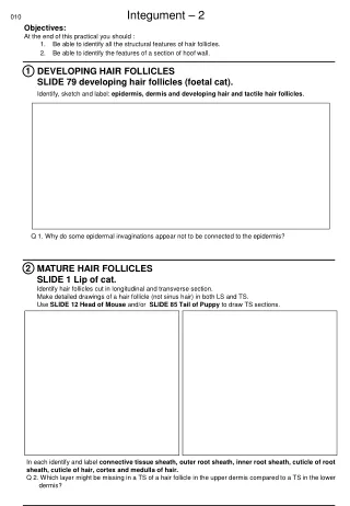
010 Integument – 2 Objectives: At the end of this practical you should : 1. Be able to identify all the structural features of hair follicles. 2. Be able to identify the features of a section of hoof wall. 1 DEVELOPING HAIR FOLLICLES SLIDE 79 developing hair follicles (foetal cat). Identify, sketch and label: epidermis, dermis and developing hair and tactile hair follicles. Q 1. Why do some epidermal invaginations appear not to be connected to the epidermis? 2 MATURE HAIR FOLLICLES SLIDE 1 Lip of cat. Identify hair follicles cut in longitudinal and transverse section. Make detailed drawings of a hair follicle (not sinus hair) in both LS and TS. Use SLIDE 12 Head of Mouse and/or SLIDE 85 Tail of Puppy to draw TS sections. In each identify and label connective tissue sheath, outer root sheath, inner root sheath, cuticle of root sheath, cuticle of hair, cortex and medulla of hair. Q 2. Which layer might be missing in a TS of a hair follicle in the upper dermis compared to a TS in the lower dermis?
SINUS HAIRS SLIDE 1 Lip of cat (or SLIDE Y sinus hair pig). Identify a sinus hair in this section. Q 3. How does the size of these hairs compare to adjacent hairs? 3 Q 4. In which hair follicle layer is the blood sinus present? Q 5. How does the sinus hair work? 4 SLIDE W skin of sheep Identify hair follicles in this section. Q 6. How do the wool hairs differ from ordinary hairs? Q 7. What are the large prominent glands in this section?
EQUINE HOOF Section of wall. Draw a low power plane of the section of equine hoof wall. Identify and label the following: Tubular horn. Inter-tubular horn. Primary lamellae of hoof. Secondary lamellae of hoof. Primary lamellae of corium. Secondary lamellae of corium. Blood vessels in corium. 5 Q 8. How would a section of bovine hoof differ from the above equine? Examine the museum specimens of equine hoof. Identify : Lamellar corium. (note, only primary lamellae will be visible as secondary lamellae are lost when separating the dermal and from epidermal components). Coronary corium with papillae of the coronary band. Perioplic (or Limbic) corium. Hoof wall. Epidermal lamellae. Sole. Frog. White line.