Integument
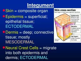
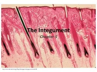
Integument. Metallic 0 Mind. Skin. Composed of. Epidermis: Stratified squamous keratinized epithelium. Dermis: Irregular collagenous connective tissue.


Integument
E N D
Presentation Transcript
Integument Metallic 0 Mind
Skin Composed of Epidermis: Stratified squamous keratinized epithelium Dermis: Irregular collagenous connective tissue The interface between them is formed by dermal ridges (papillae) that interdigitate with epidermal ridges. The two types of ridges are called rete apparatus. Hypodermis is Not from the skin it’s from superficial fascia and it’s loose connective tissue with fat
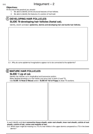
Types of Skin 1. Thick skin: • In the palms of the hand and soles of the feet. • No hair no sebaceous glands • No arrectorpilli muscles • Have sweat glands • Has the five layers of epidermis. 2.Thin skin: • Lacks stratum lucidum and stratum granulosum • Has a thin startumcorneum • Has hair, rector pilli muscles & glands
Epidermis • Keratinocytes: • Largest population. • Arranges in five layers. • Constantly renewed.
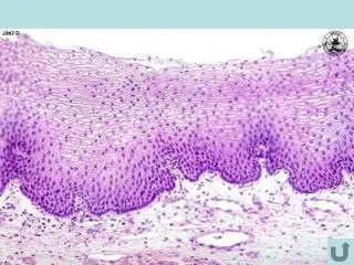
Layers of Epidermis • Stratum corneum • Stratum lucidum • Stratum granulosum • Stratum spinosum • Stratum basale
Keratinocytes migrate upward and begin to accumulate keratin filament and then they die
Stratum Basale(germinativum) • Deepest layer. • Supported by basement membranewhich separates it from dermis • Forms inter-digitations with dermis • Single layer of low columnar to cuboidal cells. • Basophilic cytoplasm and large nucleus. • Has tonofilaments • Mitoticallyactive • Have desmosomes and hemisdesmosomes
Stratum spinosum • Thickest layer. • Polyhedral to flattened cells. • Mitotically active. • With stratum basale together referred to as malpighian layer. • Rich in intermediate filaments (tonofilaments) that radiate through processes to adjacent cells forming intra-cellular bridges. • Tonofilaments tonofibrils. • Eosinophilic • Has membrane-bounded granules (lamellar granules) (contain lipid)
Stratum granulosum • 3-5 layers of flattened cells. • Most superficial layer with cells that still posses nuclei. • Has coarse granules (keratohyalin granules) • Not membrane bounded • Basophilic • Keratin filaments pass through these granules. • Has membrane bounded granules which exocytose their contents (lipids) to be a waterproof barrier.
Stratum lucidum • Clear , homogenous , slightly staining . • Present only in thick skin • Flattened thin layer of cells. • No nuclei no organelles. • Contain eleidin: Dense “packed” keratin filaments
Stratum corneum • The most superficial . • Layers of flattened , keratinized cells with thick membrane ( thick plasmalemma) • No nuclei no organelles. • Filled with keratin filaments. • The cells far from the skin surface display desmosomes. • the cells near the surface called squames (horny) cells. • They lose their connections (desmosomes) and becomes desquamated (sloughed off).
Located in stratum spinosum and dermis and oral cavity, esophagus and vagina Dense nucleus, pale cytoplasm, long processes Few mitochondria, RER, no filaments, has lysosomes Langerhans cells Has membrane bound Birbeckgranules (vermiform granules) Origin: bone marrow . Function : immune cell antigen presenting cells -> part of mononuclear phagocyte system
melanocytes • Found in stratum basale • Round to columnar cells has processes • Has oval granules (in red haired people it’s rounded) • Function produce melanin • Melanosomes contain tyrosinase enzyme that change tyrosine into melanin. • They contain melanosomes that leave melanocytes through the processes, penetrate the cytoplasm of str.spinosum cells. • Their long processes penetrate the intercellular spaces of str.spinosum.
Dermis (corium) • Dense irregular collagenous connective tissue. • Type I collagen fibers and elastic fibers. • Thicker in men • Composed of two layers: • Papillary layer • Reticular layer
Papillary layer • Loose connective tissue with • Type III collagen (reticular), elastic form networks • type VII fibers: anchoring fibers, extends from the basal lamina into the papillary layer • The C.T interdigitate with epidrmis forming dermal papillae or ridges. • Contains: fibroblast, macrophages, plasma cells, mast cells • Has capillary loops. • Has 2 mechanoreceptors: • Meissner corpuscles. • Krause end bulb
Reticular layer • Dense irregular collagenous connective tissue with Type I collagen and elastic fibers. • Rich in proteoglycan. • Contains: fibroblast, mast cells, lymphocytes, macrophages, fat cells • hair follicles , sweat & sebaceous glands..
Reticular layer (cont.) • Groups of smooth muscle fibers. • Striated muscle fibers in the face and neck also inserted in reticular layer (muscles of facial expression). • 2 mechanoreceptors: • Pacinian corpuscle. • Ruffini corpuscle.
Epidermis-Dermis Interface Interdigitation appear antiparallel on the skin as whorls, arches and loops like fingerprints (dermatoglyphs) Papillary layer forms primary dermal ridges Between the ridges are primary grooves house projections of epidermis In the center of each ridge there is secondary groove witch receives invagination of epididimisknown as interpapillarypeg, where there are dermal papillae
Sweat glands • Eccrine glands: • Located in dermis and hypodermis. • Composed of: • Simple coiled tubular glands. • Coiled duct opens on the surface of the skin at a sweat pore. • merocrine secretion.
Clear Cells • Narrow end and broad base. • No granules. • Has accumulation of glycogen. • Similar in structure with dark cells except less RER. • Watery secretion Dark Cells (mucoid cells) • Appear as inverted cone (broad end and narrow base) • RER, mitochondria, golgi apparatus, free ribosomes. • Apical granules. • Secrete mucous. Myoepithelial cells to squeeze secretion • ducts composed of stratified cuboidal epithelium.
Coiled duct : • Stratified cuboidal epithelium with 2 layers: • Cells of basal layer has large nucleus and mitochondria. • Cells of the luminal layer irregular nucleus and few organelles
Apocrine Sweat Glands They are under the influnce of sex hormones so they appear after puberty Modified apocrine glands include: Ceruminous (wax glands of external auditory canal. Glands of moll in the eyelid
Sebaceous Glands • In most of the body except: • Palms of the hands. • Soles of the feet. • Side of the feet inferior to the hairline. • Located in dermis and hypodermis. • Secretes sebum. • the ducts open in the hair follicle canal. • They are lobular with clusters of acini opening into single short ducts.
Sebaceous gland In a place with no hair follicles (Ex : lips, glans penis, areola of the nipples, labia minora, mucous surface of the prepuce) duct opens onto the surface of the skin. Under influence of hormones Holocrine secretion
Stratified squamous epithelium Composed of • Basal cells: • small • Located peripherally • Surround round cells. • Spherical nucleus. • SER, RER, glycogen and lipid droplets. • Able to divide. • Round cells: • Abundant SER. • Lipid droplets