Hypersensitivity
Hypersensitivity, or allergy, refers to inappropriate immune responses leading to harmful reactions. There are four classic types: Type I (IgE-mediated), Type II (Antibody-Mediated), Type III (Immune Complex-Mediated), and Type IV (Delayed-Type). Type I involves allergens triggering IgE antibodies, activating mast cells, and releasing mediators like histamine, which can result in conditions like asthma and anaphylaxis. Awareness of these immune responses and their management options, such as antihistamines and immunotherapy, is crucial for individuals prone to allergies.

Hypersensitivity
E N D
Presentation Transcript
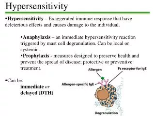
Hypersensitivity -Hypersensitivity (allergy) is an inappropriate immune response that may develop in the humoral or cell-mediated responses -Was first termed anaphylaxis -can be systematic, which often leads to shock and can be fatal, or localized, which is various atopic reactions
Types of Reactions There are four types of reactions: Type I-IgE mediated Type II-Antibody-Mediated Type III-Immune Complex-Mediated Type IV-Delayed-Type Hypersensitivity (DTH)
Type I: IgE-Mediated Hypersensitivity • hmm…that sounds bad …aren’t IgE’s supposed to be one of the 5 isotypes of “good guys”? • Of course, the allergen is the true “bad guy” – a non-parasitic antigen capable of stimulating a Type I hypersensitive response. • It’s the secretion and cross-linking of the IgE that causes the problem. • Let’s locate the IgE:
How is a Type I hypersensitive response different from a normal humoral response? • The plasma cell lineage of the B cell that exogenously processed the allergen secretes IgE, not any of the other isotypes.
What is the sequence of events in an IgE-mediated hypersensitive response? • The plasma cells secrete IgE. • These IgE bind to Fc receptors on sensitized mast cells and blood basophils. • When the allergen appears again (usually a few weeks after the first exposure), it cross-links the mIgEs and causes degranulation, releasing granules. • Mediators within these granules act on the surrounding tissues such as smooth muscle, small blood vessels, and mucous glands.
A little background on these mast cells: • Located in nearly all vascularized peripheral tissues • Contain many packets of membrane-bound granule “ammunition”, ready for release upon activation by an allergen • Can also release cytokines (thus they wear multiple immunological hats)
…and their Fc receptors… • High affinity receptor Fc,RI binds to IgE at extremely low serum concentrations (1 X 10⁻⁷). This receptor has ITAM as its cytosolic domain, thus it can initiate the process of degranulation via tyrosine phosphorylation. • Low affinity receptor Fc,RII regulates intensity of IgE response via activating B cells when cross-linked by allergen. • (check Figure 16-4 for structure details)
The chemically active effectors within the granules released via degranulation are called mediators. This group includes: • Histamines • Leukotrienes • Prostaglandins • Cytokines
Of course, these effects can be good… • Vasodilation and increased vascular permeability usher in plasma and inflammatory cells (such as esinophils, neutrophils) to attack the pathogen However, when these effects go overboard, the result is problematic: • Anaphylactic shock – extreme smooth muscle contraction compromises control of the bladder and GI tract and causes bronchiole constriction • Allergic rhinitis – excess mucous is released. More commonly known as hay fever, this ailment affects 10% of the US population • Food allergies – a variety of symptoms • Asthma – bronchoconstriction and excess mucous
Even worse for asthmatics: • They can suffer from a late-phase reaction that develops 4-6 hours after the initial reaction and persists for 1-2 days. • The late-phase reaction is caused by the heavy infiltration of inflammatory cells and the release of cytokines from mast cells, which increases the adhesion of these inflammatory cells to epithelial linings of smooth muscle. • Epithelial damage, bronchoconstriction, and inflamed bronchiole tubes results.
And just look at all these popular allergens! • What’s available in the Western medicine arsenal of drugs? • Antihistamines – block the binding of histamine on target cells • Immunotherapy – treat the patient with increased doses of the allergen (hyposensitization) to reduce severity of the response. • Or, practice better dust control, find another home for Fido, and don’t eat those strawberries!
Type II-Antibody-Mediated Cytotoxic Hypersensitivity • Involves the antibody mediated destruction of cells • Can mediated cell destruction by activating the complement system to create pores in the membrane of the foreign cell • Can also mediated by Antibody-Dependent Cell-Mediated Cytotoxicity (ADCC) where the Fc receptors bind to Fc receptor of antibody on the target cell and promote killing
Antibodies of the A,B, and O antigens are usually of the IgM class (these antigens are call isohemagglutinins) For example an A individual produce isohemagglutinins to B-like epitopes but not to A epitopes because they are self Person who are transfused with the wrong blood type will produce anti-hemmagglutinins causing complement mediated lysis Antibodies are usually of the IgG class Transfusion reactions can be delayed or immediate but have different Ig isohemagglutinins Immediate reactions has a complement-mediated lysis triggered by IgM isohemagglutinins Delayed reactions induce clonal selection and the productions of IgG which is less effective in activating the complement This leads to incomplete complement-mediated lysis Cross-matching can detect antibodies in the sera to prevent this Transfusion reactions:
Hemolytic Disease of the Newborn • This is where maternal IgG antibodies specific for fetal blood group antigens cross the placenta and destroy fetal RBC’s • Erythroblastosis fetalis-severe hemolytic disease of newborns • Most commonly develops when an Rh+ fetus expresses an Rh antigen on it’s blood that and Rh- mother doesn’t recognize
During the 1st pregnancy small amounts of fetal blood pass through the placenta but not enough to induce a responses During delivery larger amounts of fetal blood cross the placenta causing an activation of B-cells that are Rh specific thus leading to memory B-cells (anti-Rh antibodies) The IgM antibody clears the Rh+ cells from the mother In subsequent pregnancies with an Rh+ fetus, the Rh+ RBC cross the placenta activating the memory B-cells These in turn cross the placenta and damage the fetal RBC because they are seen as “foreign” This type of reaction can be prevented by administering antibodies against the Rh antigen within 25-48 hours after the 1st delivery Rhogam-is the antibody that is injected it will bind to the fetal RBC that enter the mother’s circulation and facilitate the clearance of them before B-cell activation In subsequent pregnancies the mother is unlikely to produce IgG anti-Rh antibodies If the mother doesn’t receive this injection there are other ways to treat this, depending on the severity Erythroblastosis fetalis
Drug-Induced Hemolytic Anemia • This is where certain antibiotics can absorb nonspecifically to the proteins on RBC membranes • Examples: penicillin, streptomycin • Sometimes antibodies form inducing complement-mediated lysis and thus progressive anemia • When drug is withdrawn the hemolytic anemia disappears
Type III-Immune Complex-Mediated Hypersensitivity • Reaction with antibodies create immune complexes • These generally facilitate the clearance of antigen by phagocytosis • Large amounts of immune complexes can lead to tissue damage (Type III reaction) • The magnitude depends on the quantity of immune complexes and their distribution • The complexes get deposited in tissues: • Localized reaction is when they are deposited near the site of antigen entry • When formed in the blood reaction can develop where ever they are deposited • Deposition of these complexes initiates a reaction that results in the recruitment of neutrophils • Tissue is injured by the granular release from the neutrophil (attempted phagocytosis release lytic enzymes that cause the damage)
Localized Type III Reactions: • Injection of an Antigen: • Can lead to an acute Arthus reaction within 4-8 hours • Localized tissue and vascular damage result from accumulation of fluid (edema) and RBC (erythema) • Severity can vary from mild swelling to redness to tissue necrosis • Insect bite: • May first have a rapid type I reaction • Some 4-8 hours later a typical Arthus reaction develops
Large amounts of antigens enter the blood stream and bind to antibody, circulation immune complexes can form These can’t be cleared by phagocytosis and can cause tissue damaging Type III reactions Serum Sickness-type III hypersensitivity reaction that develops when antigen is intravenously administered resulting in formation of large amounts antigen-antibody complexes and the deposition in tissue Other conditions caused by Type III- Infectious Diseases Meningitis Hepatitis Mononucleosis Drug Reactions Allergies to penicillin and sulfonamides Autoimmune Diseases Systematic lupus erythematosus Rheumatoid arthritis Generalized Type III Reactions:
Type IV Hypersensitivity a.k.a. cell mediated hypersensitivity or delayed type hypersensitivity What is delayed type hypersensitivity (DTH)? • A hypersensitive response mediated by sensitized TDTH cells, which release various cytokines and chemokines • Generally occurs 2-3 days after TDTH cells interact with antigen • An important part of host defense against intracellular parasites and bacteria
Phases of the DTH Response Sensitization phase: occurs 1-2 weeks after primary contact with Ag What happens during this phase? • TH cells are activated and clonally expanded by Ag presented together with class II MHC on an appropriate APC, such as macrophages or Langerhan cell (dendritic epidermal cell) • Generally CD4+ cells of the TH1 subtype are activated during sensitization and designated as TDTH cells
Phases of the DTH Response Effector phase: occurs upon subsequent exposure to the Ag What happens during this phase? • TDTH cells secrete a variety of cytokines and chemokines, which recruit and activate macrophages • Macrophage activation promotes phagocytic activity and increased concentration of lytic enzymes for more effective killing • Activated macrophages are also more effective in presenting Ag and function as the primary effector cell
What happens if the DTH response is prolonged? A granuloma develops… • Continuous activation of macrophages induces the macrophages to adhere closely to one another, assuming an epithelioid shape and sometimes fusing together to form giant, multinucleated cells.
Protective Role of DTH Response • A variety of intracellular pathogens and contact antigens can induce a DTH response. • Cells harboring intracellular pathogens are rapidly destroyed by lytic enzymes released by activated macrophages
Detrimental Effects of DTH Response • The initial response of the DTH is nonspecific and often results in significant damage to healthy tissue • In some cases, a DTH response can cause such extensive tissue damage that the response itself is pathogenic • Example: Mycobacterium tuberculosis – an accumulation of activated macrophages whose lysosomal enzymes destroy healthy lung tissue • In this case, tissue damage far outweighs any beneficial effects.
How Important is the DTH Response? • The AIDS virus illustrates the vitally important role of the DTH response in protecting against various intracellular pathogens. • The disease cause severe depletion of CD4+ T cells, which results in a loss of the DTH response. • AIDS patients develop life-threatening infections from intracellular pathogens that normally would not occur in individuals with intact DTH responses.