Hypersensitivity
Hypersensitivity. Department of Microbiology. Important terms. Hypersensitivity reactions are exaggerated antigen-specific immune responses which is harmful to the host. Allergen : The antigens that give rise to immediate hypersensitivity

Hypersensitivity
E N D
Presentation Transcript
Hypersensitivity Department of Microbiology
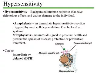
Important terms • Hypersensitivity reactionsare exaggerated antigen-specific immune responses which is harmful to the host. • Allergen:The antigens that give rise to immediate hypersensitivity • Atopy:The genetic predisposition to synthesize inappropriate levels of IgE specific for external allergens • Types of hypersensitivity: As per Coomb and Gel Classification hypersensitivity is of four types (Type I, Type II, Type III and Type IV)
Type I Hypersensitivity • Mediated by IgE antibodies. • Also known as “immediate hypersensitivity "or “Allergic response”. • Antigens that induce type I hypersensitivity are also termed as “Allergens”.
Type I Hypersensitivity • The IgE antibodies produced against Allergens remain bound to Mast cells. • Upon exposure to specific Allergens IgE antibodies induce degranulation of Mast cells. • This leads to release of inflammatory mediators like Histamine, Serotonin, Prostaglandins etc. • Some common Allergens are Pollen, Dust etc. • Example: P-K reaction
Type II Hypersensitivity • Type II Hypersensitivity is mediated by antibodies that are produced against the antigenic determinants present on cell surface. • As the antigenic determinants are present on cell surface, thus type II reaction is manifested in the form of massive cell destruction. • The target cells are lysed by: • activation of Complement cascade by the antibody molecules bound on the cell surface. • antibody dependent cell mediated cytotoxicity (ADCC). • Transfusion reactions and Hemolytic Disease of the Newborn are examples of type II reaction.
2. Mechanism of Type II hypersentivity 1. Surface antigen on target cells Target cells: Normal tissue cell, changed or modified self tissue cells Common antigen, Antigen : Blood group antigen, Drug antigen, Self-antigen modified by physical factors or infection Antigen-antibody complex 2. Antibody, complement and modified self-cell Activate complement Lyse target cells Opsonic phogacytosis Destroy target cells ADCC Mf、NK、 T Promote /surpress the target cell funcion Stimulating or blocking effect
Antigen or hapten on cell Antibody (IgG, IgM) Activate complement Opsonicphagocytosis NK , phagocyte Stimulate / block Lyse target cell Destroy target cell ADCC Target cell injury Change the function ofTarget cell Mechanism of Type II hypersensitivity
3. Common disease of type IIhypersensitivity 1)Transfusion reaction hemolysis : mismatch of ABOblood group, severely destroy RBC nonhemolysis : repeat transfusion of allogenic HLA drug anaphylactic shock:penicillin 2) Hemolytic disease of newborn Mother Rh- : first baby Rh+(Ab), second baby Rh+, fetal RBC destroyed 3) Autoimmune hemolytic anemia and type II drug reaction i. Foreign antigen or hapten Penicillin RBC hemolytic anemia QuininPlatlet thrombocytopenic purpura PyramidoneGranulocyte agranulocytosis ii. Self-antigen Drug conversion from a hapten to a full antigen induce self antibody autoimmune hemolytic anemia
Type II HypersensitivityAntibody Dependent Cell Mediated Cytotoxicity Animation: Antibodies react with epitopes on the host cell membrane and NK cells bind to the Fc of the antibodies. The NK cells then lyse the cell with pore-forming perforins and cytotoxicgranzymes
Type II HypersensitivityAntibody-Mediated Cell Disfunction Example: Myasthenia Gravis
Type III Hypersensitivity • Mediated by immune complexes (Antigen-Antibody complex). • During normal immune response only moderate quantity of immune complexes are formed and they are removed efficiently from circulation by phagocytosis. • In case of production of large quantities of immune complexes, phagocytes fail to remove all the immune complexes from circulation. • Thus, Ag-Ab complexes are deposited in various tissues. They lead to activation of complement components. Activation of complement may leads to destruction of bystander cells.
Type III Hypersensitivity • Moreover, under the influence of chemotactic complement components polymorphonuclear cells are recruited at the site. • These cells, in their attempt to engulf immune complexes, releases lysozymal enzymes in the tissue and thus cause tissue destruction. • Arthus reaction is an example of localized type III hypersensitivity. • Serum Sickness is an example of Systemic type III hypersensitivity. • Blue Eye: Dogs infected or vaccinated with live canine adenovirus I develops anterior uveitis. This lead to corneal oedema and opacity. The blue eye is considered to be an immune complexes mediated condition.
Type III Hypersensitivity • “Immune complex disease” • Soluble Ag / IgG or IgM • high titers of each required • Immune processes involved: • classical complement pathway • phagocytic cells Immunreaktionen der Haut
Soluble antigen Body Antibody Immune complex Small molecular soluble Immune complex intermediate molecular soluble Immune complex Large molecular insoluble Immune complex Deposit on the basement of capillaries Eliminate by phogacytosis Combine and activate complement system Basophils and mast cells Platelets C3a,C5a,C3b Infiltration of neutrophils Blood Clotting Mechanisms Release of vasoactive amine Phagocytose complex Release of vasoactive amine Aggregation of platlets Release the enzymes in lysosome Increase vascular permeability Increase vascular permeability Thrombus Edema Tissue injury Bleeding Edema Local or systemic immune complex diseases
3. common disease of type III hypersensitivity 1.Local immune complex disease Arthusreaction :Experimental local reaction, Necrotic vasculitis vasculitis, Ulcer Human local reaction: insulin-dependent diabetes mellitus (IDDM) 2. Acute systemic immune complex disease serum sickness Anti-serum Ab+Ag systemic tissue injury ,fever, arthritis, skin rash Pinicillin、Sulfanilamide Acute immune complex glomerulonephritis :Streptococcus infection 3. Chronic immune complex disease SLE Rheumatoid arthritis:RF+IgG Deposit on synovial membrane
Type IV hypersensitivity reaction • Because of delay in onsetof response, type IV hypersensitive reaction is also known as Delayed Type Hypersensitivity (DTH). • It approx takes 24 to 48 hours from the time of antigenic stimulation. • Unlike Type I, II, and III response (antibody mediated), Type IV reaction is mediated by Cellular immune components.
Type IV hypersensitivity reaction • The effector cells of Type IV hypersensitivity response are CD4+ (Th), CD8+ cells and activated macrophages. Some common examples of type IV hypersensitivity reaction: • Tuberculin test (used for diagnosis of Tuberculosis), • Johnin test (used for diagnosis of Johnes disease), • Mallein test (used for diagnosis of Glanders), • Brucellin test (used for diagnosis of Brucellosis).