Download

1 / 66
700 likes | 1.49k Vues
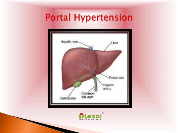
Portal Hypertension. Done by Ghassan Al- Maimani. Portal system. Conduct venous return from gut and associated organs to the liver. Much of the system is retroperitoneal but some tributaries are within mesentery. Supplies approximately 75% of hepatic blood supply.

E N D
Portal Hypertension Done by GhassanAl-Maimani
Portal system • Conduct venous return from gut and associated organs to the liver. • Much of the system is retroperitoneal but some tributaries are within mesentery. • Supplies approximately 75% of hepatic blood supply. • Portal vein is about 8 cm in length , 11 mm in diameter.
Formed at level of second lumbar vertebra by junction of the superior mesenteric & splenic veins. • Union of these veins taking place in front of IVC & behind the neck of the pancreas.
It passes upward behind the 1st part of the duodenum & then ascends in the Rt border of the lesser omentum to the Rt extremity of porta hepatis where it divides into right & left branches , which accompany the corresponding branches of the hepatic artery into substance of the liver.
Tributaries SMV Splenic vein • - Intestinal veins • Ileocolic vein • Rt colic vein • Middle colic vein • Inferior pancreatico- • douodenal vein • Rt gastro epiploic vein • IMV • Pancreatic veins • Left gastroepiploic vein • Short gastric veins. Coronary vein Para umbilical veins Cystic vein
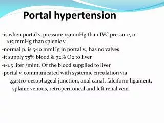
Portal HTN • It is an increase in the pressure within portal vein ≥ 12 mmHg (normal pressure 5-10mmHg)
Pathophysiology • increase in vascular resistance to the portal blood flow which determined by blood vessels radius ( small decrease in vessel radius → large increase in portal vascular resistance ). • increase in portal blood flow which is established through splanchinc arteriolar VD caused by an excessive release of endogenous vasodilators
Causes -Pre-hepatic -Intra-hepatic : presinuosoidal sinuosoidal postsinosoidal -post-hepatic
Prehepatic : • Portal vein thrombosis. • Splenic vein thrombosis. • Extrinsic compression ( tumor ). • Stenosis of portal vein. • Splanchnic arteriovenous fistula.
Intrahepatic - Presinuosoidal - Sinuosoidal & postsinuosoidal • Schistosomiasis. • Primary biliary cirrhosis. • Myeloproliferative disease. • Hepatic metastasis. • Granulomatous disease ( sarcoidosis, TB). Hepatic cirrhosis. Acute alcoholic hepatitis. Acute fulminant hepatitis. Congenital hepatic fibrosis. Vitamine A toxicity.
Post-hepatic : • Budd-chiari syndrome. • IVC obstruction. • Constrictive pericarditis. • Rt heart failure.
PortocavalAnastomosis • Esophageal anastomosis: * azygos vein (caval ) * left coronay vein ( portal ) 2. Paraumbilicalanastomosis : * epigastric veins (caval ) * parambilical veins ( portal ) 3. Rectal anastomosis : * inferior & middle hemonhoidal (caval ) * superior hemonhoidal ( portal ) 4. Retroperitoneal anastomosis : * retroperitonal parietal vein (caval ) * Visceral vein of Retzius ( portal )
History History from a patient with portal hypertension should be directed towards determining the cause of portal hypertension and, secondarily, the presence of the complications of portal hypertension.
- Determining the cause of portal hypertension involves the following: - History of jaundice - History of blood transfusions, intravenous drug use (hepatitis B and C) - Pruritus - Family history of hereditary liver disease (hemochromatosis, Wilson disease) - History of alcohol abuse
- Determining the presence of the complications of portal hypertension involves the following: - Hematemesis or melena (gastroesophageal variceal bleeding or bleeding from portal gastropathy) - Mental status changes - Increasing abdominal girth (ascites) - Abdominal pain and fever [SBP] - Hematochezia (bleeding from portal colopathy)
Examination - Signs of portosystemic collateral formation include the following: - Dilated veins in the anterior abdominal wall - Venous pattern on the flanks - Caput medusa - Rectal hemorrhoids - Ascites - Paraumbilical hernia
- Signs of liver disease include the following: • - Testicular atrophy • - Gynecomastia • - Dupuytren contracture • - Muscle wasting • - Splenomegaly - Ascites - Jaundice - Spider angiomas - Palmarerythema • Asterixis
- Signs of hyperdynamic circulatory state include the following: - Bounding pulses - Warm, well-perfused extremities - Arterial hypotension
Workup Lab Studies: • Lab studies are directed towards investigating etiologies of cirrhosis, which is the most common cause of portal HTN • - Antinuclear antibody, antimitochondrial antibody, antismooth muscle antibody • - Iron indices • - Alpha1-antitrypsin deficiency • - Ceruloplasmin, • - Liver function tests • - Prothrombin time • - Albumin • - Viral hepatitis serologies • - Platelet count
Imaging Studies: - Duplex-Doppler ultrasonography - Ultrasound (US) is a safe, economical, and effective method for screening for portal hypertension. - Features suggestive of hepatic cirrhosis with portal hypertension include the following: • Nodular liver surface is suggestive. • Splenomegaly is a suggestive finding. • Patients may demonstrate the presence of collateral circulation.
- CT scan :- - CT scan is a useful qualitative study in cases where sonographic evaluations are inconclusive. - Findings suggestive of portal hypertension include the following: • Collaterals arising from the portal system are suggestive of portal hypertension. • Dilatation of the IVC also is suggestive of portal hypertension.
- Magnetic resonance imaging - MRI provides qualitative information similar to CT scan when Doppler findings are inconclusive. - Liver-spleen scan • - This is described for historical interest only. • - Liver-spleen scan uses technetium sulfur colloid taken up by cells in the reticuloendothelial system. • - A colloidal shift from the liver to the spleen or bone marrow is suggestive of increased portal pressure.
Other Tests: • Selective angiography of the superior mesenteric artery or splenic artery with venous return phase.
Procedures: Hemodynamic measurement of portal pressure - The most commonly used method is measurement of the hepatic venous pressure gradient (HVPG), which is an indirect measurement that closely approximates portal venous pressure. - A fluid-filled balloon catheter is introduced into the femoral or internal jugular vein and advanced under fluoroscopy into a branch of the hepatic vein. Free hepatic venous pressure (FHVP) then is measured. The balloon is inflated until it is wedged inside the hepatic vein, occluding it completely, thus equalizing the pressure throughout the static column of blood. The occluded hepatic venous pressure (ie, wedged hepatic venous pressure) minus the unoccluded, or free, portal venous pressure (ie, FHVP) is the HVPG.
Endoscopy • Perform upper endoscopy, as appropriate, to screen for varices in every patient with suggestive findings of portal hypertension. • Gastroesophageal varices confirm the diagnosis of portal hypertension; however, their absence does not rule it out. • Various indirect indices, such as platelet count, spleen size, albumin, and Child-Pugh score, have been studied to help diagnose varices without endoscopy.
- A recent case review study revealed some of these predictors as unreliable. For the time being, endoscopy remains the criterion standard for screening patients with cirrhosis for varices. • In compensated patients without varices, repeat endoscopy at 2- to 3-year intervals to evaluate for the development of varices. • - In compensated patients with small varices, repeat endoscopy at 1- to 2-year intervals to evaluate the progression of varices.
Child-Pugh score -The Child-Pugh classification is used to assess the prognosis of chronic liver disease, mainly cirrhosis - It consists of 2 parts : - laboratory : bilirubin and albumin - clinical : encephalopathy , ascites, PT
Treatment • Gastroesophageal variceal hemorrhage is the most dramatic and lethal complication of portal hypertension; therefore, most of the following discussion focuses on the treatment of variceal hemorrhage - General measures - Medical - Endoscopic - Surgical
General measures • admit to ICU. • obtain iv access. • resuscitation: prompt but do not overdo it. Blood : Aim Hb = 8 g/dl NS • FFP for coagulopathy ( bring INR < 1.4 ) • Platelets for sever thrombocytopenia ( bring PLT > 50,000) • Intubation-prior to endoscopy ( for airway prophylaxis).
Medical • prophylactic Abx :- • Improve survival ( meta-analysis ) • rebleeding. • infections. • Norfloxacin 400 mg po bid 7 days.
Vasoactive agents :- • Splanchnic VCs. • reduce inflow into portal vein. • somatostatin • + analogues :- octreotide. • vasopressin • + analogues :- terlipressin.
octreotide • No effect on mortality. • reduce rebleeding when added to endoscopic therapy. • 50 mcg/h bolus, 3-5 days.
vasopressin • Very potent splanchnic VC • Unaceptable side effects :- - cardiac ischemia. - peripheral ischemia. - Bowel ischemia. - arrhythmia.
Nonselective B-blockes :- • reduce portal and collateral blood flow • Splanchnic vasoconstriction • Propranolol is administered at a dose of 20 mg every 12 h • A recent meta-analysis of 11 trials evaluating nonselective beta-blockers in the prevention of first variceal bleeding shows that the bleeding rate in controls (25%) is significantly reduced (to 15%) in patients treated with beta-blockers after a median follow-up of 24 months • Beta-blockers are best continued for the patient's lifetime because the risk of variceal hemorrhage returns to that of the untreated population once beta-blockers are withdrawn
Endoscopic Rx • Perform within 12 h • After resuscitation / intubation . • band ligation is better than sclerotherapy for control of bleeding and rebleeding.
Endoscopic banding • Occludes venous channels. • Multiple sessions + surveillance. • > 60 % rebleed. • 13 fail Rx. • Less complication Vs sclerotherapy Rx. • Endoscopic Rx of choice.
Endoscopic Sclerotherapy • Intra- or Para- Variceal • Occludes venous channels • Multiple sessions + surveillance • >60% rebleed • 1/3 fail treatment • 30% complication rate
Endoscopic Sclerotherapy Intravariceal Paravariceal
Complications of ScleroTx • LOCAL • Ulceration • Stricture • Perforation • SYSTEMIC • Fever • Pneumonitis • CNS
Other intervention • Balloon-tube tamponade • The Minnesota tube • The Minnesota tube is an adaptation of the Sengstaken-Blakemore (S-B) tube, the difference is that the S-B tube does not have the esophageal suction port to prevent aspiration
Surgical options • Total shunt. • Selective shunt. • Partial shunt. • Non-shunt.
Total Shunts End to Side Portocaval Side to Side Portocaval Interposition Shunts Central Splenorenal
Total Shunt Results • Prevent rebleed > 90% • Thrombosis with graft • Encephalopathy rate 40%
Selective Shunts • Goals: • Prevent variceal bleeding and encephalopathy • Mechanism: • Decompress Varices • Maintain Portal Perfusion • Maintain Portal Hypertension • Key: • Decompress onlygastrosplenic compartment