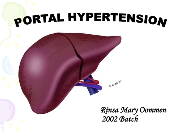
Portal hypertension
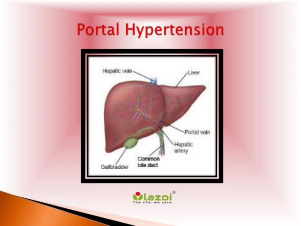
Portal hypertension. - is when portal v. pressure >5mmHg than IVC pressure, or >15 mmHg than splenic v. -normal p. is 5-10 mmHg in portal v., has no valves -it supply 75% blood & 72% O2 to liver -1-1.5 liter /mint. Of the blood supplied to liver

Portal hypertension
E N D
Presentation Transcript
Portal hypertension -is when portal v. pressure >5mmHg than IVC pressure, or >15 mmHg than splenic v. -normal p. is 5-10 mmHg in portal v., has no valves -it supply 75% blood & 72% O2 to liver -1-1.5 liter /mint. Of the blood supplied to liver -portal v. communicated with systemic circulation via .gastro-oesophageal junction, anal canal, falciform ligament, splanic venous, retroperitoneal and left renal vein.
Etiology of portal hypertension:- -most common cause is liver cirrhosis -it may be :- 1- presinusoidal 2- sinusoidal 3-postsinusoidal
Clinical features:- 1-haematemesis duo to gastro-esophageal varices 2-splenomegaly associated with hypersplenism causing pancytopenia 3-caput medusa duo to umbilical vein opening, you may hear audible venouse hum( Cruveilhier_ Baumgarten murmur) 4-ascites 5-anorectal varices 6-increasing cardiac out put causing generalised vasodilatation.
1-Treatment of esophageal varices:- A-prevention of bleeding .abstention from alcohol .avoidance of aspirin, NSAID .B-blokers as propranolol, nadolol .prophylactic endoscopic variceal ligation
B-Treatment acute variceal bleeding 1-admition ICU 2-blood resuscitation 3-coagulopathy problems , FFP , plasmin, vit K injection 4-cirrhotic pts. High risk for bacterial infection, short term prophylaxis as Ceftriaxone 5-drugs Vasopressin or somatostatin or its analogue 6- octerotides 7- oesophagogastroduodenoscopy (OGD) .sclerotherapy with ethanolamine oleate .banding 8-10-20% needs shunt treatment or TIPSS (TransjugularIntrahepaticPortosystemic Stent Shunt ) 9-ballon tamponade ( Sengstaken-Blakemore tube
2-Treatment of gastric varicesis the same lines as esophageal varices 3-Surgical shunts. which is decreased since introduction of the TIPSS and liver transplantation. 4-treatment of cases duo to portal vein & splenic thrombosis by Sugiuraoperation ( splenectomy ,lower esophagouse and gastric devasculation.
Ascites -accumulation of free peritoneal fluid -common features of the advanced liver disease independent of the etiology -Etiology:- 1-advanced liver disease 2-cancer 3-acute pancreatitis ascitis 4-TB 5-primary peritonitis 6- other causes
Clinical features:--insidious-abdominal discomfort and dragging sensation Diagnosis:- -clinically -u/s and CT-abdomen confirm it -aspiration .cytology .amylase content .C&S test
Treatment:- 1-restrict additional salt intake 2-diuretics as frusemide, spironolactone 3-prevent liver impairment by avoiding precipitating factors as alcohol 4-avoid hypo-natremia and hypo-kalemia 5-abdominal paracentesis 6-liver transplantation for ascites in diuretic resistant ascites and patients with deterioration liver v function ( rising bilirubin, dropping albumin, prolong PT ) 7-peritoneovenouse shunting ( Le Veen shunt, Denver shunt) 8-TIPSS
Benign liver lesions Include:- 1-cysts 2-Haemangioma 3-adenoma 4-focal nodular hyperplasia 5-bile duct hamartoma
1-Hemangioma -most common benign lesion, 2-20% of the population -common in women -consist of abnormal plexus of blood vessels, endothelial -lined space, contain fibrous tissue, congenital. -size range from < 1 to 10-25 cm. called giant cavernous hemangioma -often multiple -clinical features:- -pain -rarely rapture spontaneously to peritoneal cavity -diagnosis:- - by U/S, if uncertain do CT abdomen. -no percutaenouse biopsy …..bleeding occur -treatment:- -small one no treatment -if symptomatic ( pain) …surgery ( enucleation or formal resection) -if giant type ….controversial
2-Hepatic adenoma -rare -common in young women -well circumscribed& vascular solid tumor with no bile duct gland or kuffer cells. -malignant risk potentially to change to hepatocellular carcinoma (HCC) -clinically the patient may have abdominal pain or bleeding because it caries also significant risk of spontaneous rupture with intra-peritoneal bleeding. -associated with sex hormones including oral contraceptive pill(OCP) -regression of symptomatic adenoma occur if pt. stop using pill. -radiologically can not differentiate from malignant lesions, so do CT-Abdomen angiography -confirm the diagnosis by liver biopsy -treatment is resection
3-Focal nodular hyperplasia:- -unusual benign lesion of unknown etiology -no association with underlying liver disease -common in middle aged women -it is a focal overgrowth of the functioning liver tissue supported by fibrous stroma, with central scar tissue. -do not rapture and has also no significant change to carcinoma. -may cause abdominal pain. -diagnosis by U/S- CT-abdomen but sulpher colloid liver scanning differentiate it from benign adenoma and primary or metastatic cancer lesions. -treatment if causing pain do resection and stop using pill.
4-Bile duct hamartoma:- -small lesions (2-4mm) on surface of the liver. -firm, smooth, whitish-yellow in colure -difficult to differentiate it from metastatic cancer -Treatment needs excisional biopsy
Malignant liver tumors Include:- 1-Primary .Hepto-cellular cancer (HCC) .Cholangio-carcinoma(bile duct cancer) .Gal bladder cancer 2-Metastatic .Colorectal metas, is commonest cause for metas., 60% metastases to liver .Neuro-endocrine cancer( carcinoid, islet cell tumor) .Other metastatic tumors, nearly every cancer has propensity to metas. to liver specially breast, long, other GIT tumors
Hepato-cellular carcinoma -5th commonest carcinoma worldwide -1 000 000 new cases annually -increase incidence duo to association with chronic liver disease (HBV,HCV ) -so screening of patients with viral disease with serial liver sonography and AFP is necessary -multifocal usually -middle age affected mostly -70-80 % has liver cirrhosis -in cirrhosis 3-6% annually change to HCC
Hcc cont. -metastases to long and bone, so needs CT of the chest and bones for staging especially -risk factors:- -HBV,HCV -Alcoholic cirrhosis -hemochromatosis -nonalcoholic steatohepatitis -Clinical features:- symptoms of chronic liver disease ( malaise, weakness, jaundice, ascites, variceal bleeding, encephalopathy) or anorxia, lose of weight in advanced cancer.