Download
1 / 51
800 likes | 1.85k Vues
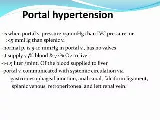
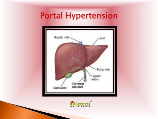
Portal Hypertension. Portal vein anatomy. The portal vein is formed in front of IVC and behind the neck of the pancreas ( at the level of 2 nd lumber vertebra ) by union of the splenic & SMV. It is 7-8 cm in length & contains no valves.

E N D
Portal vein anatomy • The portal vein is formed in front of IVC and behind the neck of the pancreas ( at the level of 2nd lumber vertebra ) by union of the splenic & SMV. • It is 7-8 cm in length & contains no valves. • It courses in the lesser omentum posterior to both the common hepatic artery & common bile duct.
Causes of portal hypertension • Increased resistance to flow A)Pre-hepatic (portal vein obstruction): • 1- congenital atresia or stenosis. • 2- thrombosis of portal vein. • 3- thrombosis of splenic vein. • 4- Extrinsic compression (e.g , tumor).
Causes of portal hypertension B)Intra-hepatic: • 1- liver cirrhosis obstruction is sinusoidal & post-sinusoidal. • 2- bilharzial periportal fibrosis obstruction is pre-sinusoidal.
Causes of portal hypertension The causes of portal hypertension in cirrhotic patients are: • Diminution of the total vascular bed by obliteration, distortion, & compression of sinusoids. • Compression of the tiny radicals of portal & hepatic veins by excessive fibrosis. • Development of multiple arteriovenous shunts between the branches of the hepatic artery & portal vein.
Causes of portal hypertension C) Post-hepatic: • 1- Budd-Chiari syndrome ( hepatic vein thrombosis ) prominent ascites ,hepatomegaly & abdominal pain. • 2- Veno-occlusive disease. • 3- Cardiac disease: a- constrictive pericarditis. b- valvular heart disease. c- right heart failure.
Causes of portal hypertension • Increased portal blood flow A)Arterial-portal venous fistula. B)Increased splenic flow: 1- Banti’s syndrome ( liver disease secondary to splenic disease, result from cirrhosis & other hepatic disorders) 2- Splenomegaly (e.g tropical splenomegaly ,hematologic diseases).
Sequelae & Clinical picture 1-Porto-systemic collaterals: • In normal conditinos collapsed. • In portal hypertension engorged divert blood away from the portal circulation.
Sequelae & Clinical picture • The important sites of these collaterals are: a)At the lower end of oesophagus Oesophageal tributaries of Lt gastric vein (portal) Oesophageal tributaries of hemiazygous vein (systemic). b)Around the umbilicus Para umbilical vein (portal) Superior & inferior epigastric veins (systemic)
Sequelae & Clinical picture c)Lower rectum & analcanal Superior rectal vein (portal) Middle & Inferior rectal veins (systemic) d)At the back of the colon Rt & Lt colic veins (portal) Rt & Lt renal veins (systemic) e)Retroperitoneum Tributaries of superior & inferior mesentric veins { Retzius } (portal) Posterior abdominal & subdiaphragmatic veins (systemic)
Sequelae & Clinical picture 2- Splenomegaly • The most constant physical finding. • In 80% of patients regardless the cause. * In Bilharzial cases : • at 1st due to reticuloendothelial hyperplasia due to absorption of bilharsial toxins. • With progress of portal hypertension due to congestion.
Sequelae & Clinical picture 3-Congestion of the whole GIT • Leads to anorexia, dyspepsia, indigestion, and malabsorption. 4-Bleeding varices. 5-Ascites (multifactorial) • Portal hypertension alone cannot cause ascites. • Hypoalbuminaemia below 3 gm/100 ml. • Salt &water retention high level of aldosterone, oestrogens & anti-duretic hormone. • Increased lymphatic transudation from liver surface.
Investigations 1.Assessment of liver function tests • (a)Hypoalbuminaemia. The liver is the only site of albumin synthesis. • (b)ALT & AST are moderately raised. • (c)Prothrombin time and concentration are disturbed. This test is the most sensitive liver function.
Investigations 2.Detection of oesophageal varices by: (a)Fibreoptic upper endoscopy (b)Barium swallow can visualize varices in 90% of cases. They appear as multiple, smooth, rounded filling defects (honey-comb appearance) (c)Duplex scan can show dilated portal vein and collaterals.
Investigations 3.Detection of splenic sequestration and hypersplenism. • (a) Blood picture anaemia, leucopenia, thrombocytopenia or pancytopenia. • (b) Bone marrow examination hypercellularity. • (c) Radioactive isotope studies : using the patients own RBCs tagged with 51Cr diminished half life of RBCs & increased radioactivity over the spleen.
Investigations 4.Diagnosis of the aetiology of liver disease is performed by: • (a) Immunological tests for hepatitis markers. • (b) Liver biopsy after assessment of prothrombin time and concentration.
Child Pugh classification Grade A, 5-6 points; Grade B, 7-9 points; Grade C, 10-15 points
Treatment Management of patients with actively bleeding oesophageal varices 1.Admission. The patient should be admitted to hospital. 2.Resuscitation. • A wide bore cannula is inserted. • A blood sample is taken. • Restoration of blood volume should be rapid. • Overexpansion of the circulation should be avoided . • Morphine and Pethidine are contraindicated.
Treatment 3.Correct coagulopathy. • Vitamin K is administered intravenously. 4.Prevent encephalopathy. Blood in the intestine will be fermented to ammonia and other nitrogenous products. • Repeated enemas. • Oral lactulose. This is a disaccharide sugar, fermented by the intestinal flora lactic acid combines with ammonia. • Neomycin 0.5 gm every 4 hours can reduce the bacterial flora.
Treatment Sclerotherapy • Intra- or Para- Variceal. • 1-3 ml sclerosant (ethanolamine oleate). • Occludes venous channels. • Multiple sessions (2 weekly). • Control bleeding in 80-95 %. • About 50% rebleed. • 30% complication rate.
Endoscopic Sclerotherapy Intra-variceal Para-variceal
LOCAL Ulceration. Stricture. Perforation. Retrosternal discomfort for few days. SYSTEMIC Fever Pneumonitis CNS Complications of Sclerotherapy
Treatment Endoscopic Banding • Occludes venous channels • Sessions < sclerotherapy • Same results as sclerotherapy • complications vs sclerotherapy • Endoscopic treatment of choice
Treatment Drugs Vasopressin vasoconstriction of the splanchnic circulation. Disadvantages • colicky abdominal pains, & diarrhoea . • anginal pains, so it is contraindicated in the elderly. • Produce temporary control of bleeding in about 80% of cases. • To prolong its action it is combined with glycine (Glypressin).
Treatment Somatostatin • lower the intravariceal pressure without significant side effects. • Initial bolus 100 microgram continuous infusion of 25 microgram/ h for 24 hs. Beta blockare bleeding by cardiac output. Does 20-60 mg bid 25% in HR. Reduces 40% of bleeding episodes Does not reduce mortality
Treatment Balloon tamponade by Sengestaken or Linton tube. • The gastric balloon is inflated first by 200 ml of air, and pulled upwards to press the gastric fundus. • If bleeding continues, the oesophageal balloon is inflated. • The pressure in the oesophageal balloon should not exceed 40 mm Hg. • This therapy is effective in controlling bleeding in 80-90% of cases.
Treatment Disadvantages : • Discomfort to the patient. • The patient cannot swallow his saliva • Liability to cause oesophageal ulceration or stricture. • Once the tube is deflated, there is liability to rebleeding in 60-80% of patients. Balloon tamponade is only used as a temporary measure before sclerotherapy or surgery.
Treatment Emergency surgery. If all the previous measures fail to stop bleeding, surgery is recommended. • If the general condition of the patient is satisfactory splenectomy, portoazygos disconnection and stapling of the oesophagus. • If the patient is not very fit stapling alone can be performed.
Treatment • Trans-juguJar Intra-hepatic Porto-Systemic Shunt ( TIPSS )
Treatment Indications for TIPSS: • Refractory bleeding • Prior to transplant • Child C • Refractory ascites Main early complication: • Perforation of liver capsule massive haemorrhage.
Treatment Treatment of patients with history of bleeding oesophageal varices: • 1. Repeated sclerotherapy until the varices are obliterated is the first choice. • 2. Elective surgery is mainly indicated if sclerotherapy failed to stop recurrent attacks of bleeding provided that they are fit.
Treatment Operations for portal hypertension Shunt operations. • The idea of these operations is to lower the portal pressure by shunting the portal blood away from the liver
Total shunt operations 1-Porta-caval operation End to side Side to side
Porta-caval operation • very efficient in lowering the portal pressure no bleeding occurs from the varices. disadvantages: • deprives the liver of portal blood flow accelerates the onset of liver failure. • Recurrent hepatic encephalopathy in 30-50% of patients.
Proximal spleno-renal shunt • indicated if the portal vein is thrombosed or if splenectormy is indicated due to hypersplenism . • The incidence of encephalopathy is less than after porta caval shunt. • it is less effective In preventing further bleeding. • If the splenic vein is less than 1 cm the anastmosis is liable to thrombosis.
Mesocaval (Drapanas) shunt • insertion of a a synthetic graft as dacron, or autogenic vein between the superior mesenteric vein and inferior vena cava. • The incidence of thrombosis is high
Selective shunt (Warren shunt) • The Rt and Lt gastric vessels are ligated. • The proximal end of splenic vein is ligated while the distal end is anastomosed to the left renal vein. • The short gastric veins are preserved and will selectively decompress the lower end of the oesophagus. • The incidence of encephalopathy is low, and the liver functions remain normal.
Porta azygos disconnection operations There are many techniques for performing devascularization. Hassab Khairy operation • splenectomy & ligation of the Rt and Lt gastric vessels, the short gastric vessels and the vascular arcade along the greater curvature of the stomach leaving only the right gastroepiploic vessels.
All vessels surrounding the lower 5-10 cm of the oesophagus are ligated. • There is no encephalopathy following this operation and the portal blood flow is intact. • There is a low incidence of rebleeding following the operation, but it can usually be controlled by sclerotherapy.
Liver Transplant • Indicated for liver failure Not for variceal bleeding. • 24% in USA die on waiting list
Control of Ascites • Sodium / Water Restriction. • Spironolactone. • Loop Diuretic. • Large Volume Paracentesis. • Peritoneal-Venous Shunt . • TIPSS
Splenic Vein Thrombosis • Etiology: Pancreatitis - Acute or Chronic Pancreatic Carcinoma • Hallmark: Isolated Gastric Varices • Treatment: Splenectomy (if bleeding)
Portal Vein Thrombosis Etiology: Congenital - “Cavernous Transformation” Hallmark: Normal Liver Function W/ Varices Treatment: Endo Tx OR DSRS
Budd-Chiari Syndrome • Etiology Hypercoagulable: Estrogens, XRT, Myeloprolif, PNH IVC Occlusion: RA Myxoma, Pericarditis, Liver Mass High Dose ChemoTx • Presentation: Classic Triad Abdominal Pain Ascites Hepatomegaly