Download
1 / 18
220 likes | 824 Vues
Central nervous system (CNS) Brain + Spinal Cord. Forebrain telencephalon cortex, basal ganglia diencephalon thalamus, hypothalamus Midbrain tectum, tegmentum Hindbrain cerebellum, pons, medulla. cerebral hemispheres. brain stem. Cerebral cortex. central sulcus. postcentral gyrus.

E N D
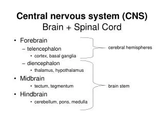
Central nervous system (CNS) Brain + Spinal Cord • Forebrain • telencephalon • cortex, basal ganglia • diencephalon • thalamus, hypothalamus • Midbrain • tectum, tegmentum • Hindbrain • cerebellum, pons, medulla cerebral hemispheres brain stem
Cerebral cortex central sulcus postcentral gyrus • Surface is folded to increase area: • Sulcus (pl. sulci) = groove • Gyrus (pl. gyri) = bulge between sulci • Grey matter = cell bodies • White matter = axons grey matter white matter
Cerebral hemispheres • Left & right hemispheres are connected by white matter tracts called commissures. • These allow communication between lateralized brain areas. • Largest commisure is the corpus callosum. • In “split brain” patients the corpus callosum is transected, leading to neuropsychological deficits. corpus callosum
Lobes of the cerebral cortex parietal lobe -body sense -multimodal integration frontal lobe -motor -executive functions occipital lobe -visual temporal lobe -memory -auditory
Primary & association cortex • Primary cortical areas are most directly linked to the sensory or motor systems in the body. • These areas project to association cortex, allowing integration of information. • Association cortex is where sophisticated, higher-level processing takes place, e.g.: • planning of a sequence of movements - motor association cortex, frontal lobe • perceiving a visual object - visual association cortex, temporal lobe • making decisions - multimodal association cortex, frontal lobe
Primary cortical areas primary somatosensory area primary motor area medial surface of right hemisphere central sulcus primary visual area lateral fissure (Sylvian fissure) lateral surface of left hemisphere primary auditory area
Functional Distribution of Cortex 1. Primary Cortex (Direct Projection Areas) • 3 Sensory—Visual, Auditory, Somatosensory— • 1 Motor. 2. Secondary Cortex (unimodal) 3. Tertiary Cortex (Association, polymodal)
Subcortical structures • In the forebrain, these are: • in the telencephalon, the basal ganglia and the limbic system. • in the diencephalon, the thalamus and hypothalamus. basal ganglia thalamus hypothalamus
Basal ganglia • important in motor control and cognition. • Damage to the basal ganglia occurs in Parkinson’s disease and Huntington’s disease. • 5 Components: • 1. Caudate Nucleus • 2. Putamen • 3. Globus Pallidus • 4. Subthalamic Nucleus • 5. Substantia Nigra
Limbic system cingulate gyrus • Functions include emotion and memory. • Limbic system includes cortical & subcortical structures: • Cingulate gyrus (cognitive control). • Hippocampus, fornix & mamillary bodies (episodic memory). • Amygdala (emotion). hippocampus amygdala
Diencephalon • Thalamus: • Closely connected with cerebral cortex & its functions. • Thalamic nuclei have distinct functions, e.g. lateral geniculate nucleus in vision. • Hypothalamus: • Controls autonomic nervous system and endocrine system (hormones). • Last sensory way station on the way to the cortex.
Thalamic nuclei • 1. Ventral Posterior Nuclei: Somatosensory • 2. Lateral Geniculate: Visual • 3. Medial Geniculate: Auditory • Pulvinar connects with parietal lobe and is a major part of an attentional control system.
Midbrain tegmentum • Midbrain (and hindbrain) structures perform relatively primitive functions, e.g. reflexes. • Tectum comprises: • superior colliculi, fish’s visual system • inferior colliculi, fish’s auditory system. • Tegmentum includes nuclei involved with : • arousal (reticular formation) • species-specific behavior (periaqueductal grey) • motor control (red nucleus, substantia nigra) tectum
Hindbrain • Cerebellum • important for precise movement control and learning. • also involved in cognition. • Pons: • nucleus relays info. from cortex to cerebellum. • contains reticular formation - arousal. • Medulla (oblongata): • necessary for vital functions: breathing, heartbeat. pons cerebellum medulla
BRAINSTEM • Many neurotransmitter systems • Reticular Activating System • Sleep & wakefulness control • Controls respiration and other bodily functions • Midbrain • Pons • Medulla
Brainstem Components • Midbrain • Superior Colliculus: Eye movements and visual reflex functions. • Inferior Colliculus: Auditory reflex functions. • Pons & Medulla. • Ascending sensory and descending motor pathways. Bulge of Pons caused by pathways to the cerebellum.
Cerebellum • Inputs from sensory & motor centers and vestibular system. • Outputs to spinal cord and thalamus (then cortex). • Functions: • Maintenance of posture, and fine motor control (timing). • Also involved in higher functions including language