Mitochondrial Disorders
Mitochondrial Disorders. Chenjie Xia (PGY-IV) Montreal Neurological Institute Wednesday, Jan. 25 th , 2012. Outline. Basic mitochondrial molecular biology Clinical implications of mitochondrial genetics Clinical approach to mitochondrial disorders General features

Mitochondrial Disorders
E N D
Presentation Transcript
Mitochondrial Disorders Chenjie Xia (PGY-IV) Montreal Neurological Institute Wednesday, Jan. 25th, 2012
Outline • Basic mitochondrial molecular biology • Clinical implications of mitochondrial genetics • Clinical approach to mitochondrial disorders • General features • Visual loss, ophthalmoplegia, peripheral neuropathy, ataxia
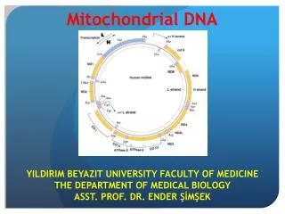
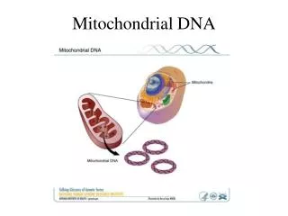
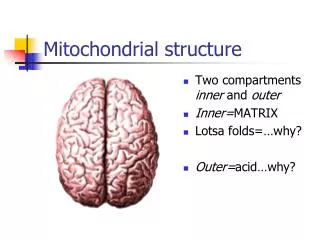
Mitochondrion Basics • 4 compartments: • outer mb • inner mb (folds into cristae) • intermembrane space • matrix Larsson and Oldfors, Acta Physiol Scand 2001.
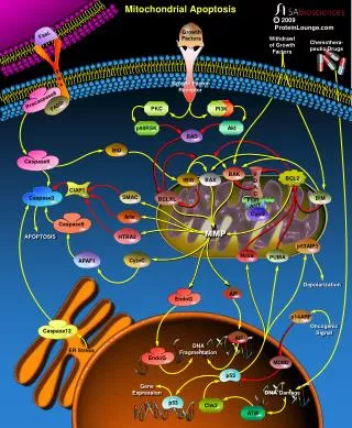
Role of mitochondria = energy production Berardo et al. Curr Neurol Neurosci Report, 2010
Respiratory Chain http://www.photobiology.info/Hamblin.html
Mitochondrion Basics • Mitochondrial respiratory chain: • Most “lucrative” step for ATP production • 5 enzymes complexes (90 protein subunits) • complexes I, II, III, IV creates proton gradient (pump protons out of matrix) • complex V uses H gradient to generate ATP • Mobile electron carriers: CoQ, cyt c
Mitochondria Genetics • mt components activity depend on: • nuclear DNA (nDNA) • mitochondrial DNA (mtDNA) Larsson and Oldfors, Acta Physiol Scand 2001.
Mitochondrial Genetics • mt genome encodes only 37 genes • 13/90 proteins of the RC • 2 rRNAs, 22 tRNAs • Most mt proteins encoded by nDNA • e.g. complex II, CPTII, PDC • nDNA exerts +++ control on mt DNA & proteins • Concept of intergenomic communication • Replication of mtDNA depend on factors encoded by nDNA
Mitochondrial Genetics • nDNA mutations • Usually manifest in childhood • More severe and diffuse • mtDNA mutations • Usually manifest in adulthood • More indolent and mosaic • These principles hold less well given recent discoveries showing increasing clinical and genetic heterogeneity of mt disorders
Mitochondrial Genetics • Concept of heteroplasmy • Mitochondrion contain mix of mutant and wild-type mtDNA • Proportion of mutant mtDNA differs in different tissues or even cells of same tissue • Concept of threshold effect • Sx develop only when mutant mtDNAreaches certain threshold (usually high, >90%) • Threshold depends on energy metabolism of tissue • Concept of replicative segregation • Mutant mtDNA are “selected out” with repeated mitoses, but accumulate in tissues not undergoing mitoses (e.g. neurons, muscles)
Mitochondria Genetics • Mitochondrial disorders can be sporadic or inherited • If inherited, mainly maternally: • “Bottle-neck” effect in oogenesis • rare case report of paternally inherited • Often no clear genotype-phenotype correlation
Mitochondrial Genetics • THM =phenotypic expression of mt disorders depend on many factors: • Nuclear versus mitochondrial mutation • Pathogenicity of mutation itself • Heteroplasmy • Threshold effect • Mitotic activity of tissue • Energy demand of tissue • Age • Etc…
General features of MID • Classic S/Sx: • Neurological: • stroke, seizure, dev. delay, dementia, visual impairment, EOMs, deafness, neuropathy, myopathy • Other: • DM, hepatopathy, cardiomyopathy, cardiac conduction defects, short stature
Clinical Manifestations - Systemic Nardin and Johns. Muscle and Nerve, 2001.
General features of MID • Key points: • +++ multisystemic • +++ overlap b/w different syndromes • same mutation can cause different phenotypes • proportion of 3243tRNA mutation determines CPEO vs MELAS vs Leigh’s • same phenotype can result from different mutations • MELAS can result from 3243tRNA, 3271tRNA, 11084 ND4
Classification of MIDs • 1.Affected structure or pathway w/i mito. • RC subunits, tRNA, mt transport machinery, mt maintenance, etc • 2.Mono- vs multi-systemic • 3. Syndromic vs non-syndromic • +++ genetic & phenotypic overlap b/w the two • Syndromic better known for acronyms and for understanding of mito medicine • But non-syndromic more common, probably less recognized in clinical practice (atypical & less spectacular presentations)
Presenting Phenotypes • Visual Loss • Ptosis / opthalmoplegia • Neuropathy • Ataxia • (Myopathy) • (Seizures) • (Stroke)
Genetic Defects of MIDs Finsterer, CJNS, 2009
Presenting Phenotypes • Visual Loss • Ptosis / opthalmoplegia • Neuropathy • Ataxia • (Myopathy) • (Seizures) • (Stroke)
Visual Loss – LHON • Leber’s Hereditary Optic Neuropathy • Degeneration of retinal ganglion cells • Most common disease caused by mtDNA mutation • Clinical presentation • Bilateral sequential acute or subacute visual failure • Central vision lost before peripheral, blue-yellow perception lost early on (red-green more preserved) • Disc swelling and hyperemia followed by atrophy • Predominantly in young men • Little or no recovery (altho visual impairment seldom complete)
Visual Loss = LHON • When to think of LHON for visual loss: • Young men • no vascular comorbidities (less likely ischemic) • Painless (less likely optic neuritis, either viral or demyelinating) • No toxic or deficiency state (B12, thiamine, tobacco-alcohol amblyopia, sildenafil)
Genetic Defects of MIDs Finsterer, CJNS, 2009
Visual Loss - RP • Retinitis Pigmentosa • All retinal layers affected • Predominance in males • 1st Sx = nyctalopia (impairment of twilight vision) • Usu both eyes affected simultaneously • Perimacular zones affected first partial to complete ring scotoma • Pigmentary changes spare fovea eventually pt perceives world as if seeing through tubes
Visual Loss - RP • DDx • Bardet-Biedl syndrome, Laurence-Moon syndrome, Freidreich’s ataxia, Refsum, Cockayne syndrome, Bassen-Kornzweig disease • Kearn-Sayre syndrome
Presenting Phenotypes • Visual Loss • Ptosis / opthalmoplegia • Neuropathy • Ataxia • (Myopathy) • (Seizures) • (Stroke)
Ophthalmoplegia – KSS • Kearns-Sayre syndrome • Obligatory triad: onset before 20, pigmentary retinopathy, progressive external opthalmoplegia • Other features:cardiac conduction abnormalities, can also have high CSF protein, cerebellar ataxia, seizures, sensorineural deafness, pyramidal signs
Genetic Defects of MIDs Finsterer, CJNS, 2009
Ophthalmoplegia – CPEO • Chronic progressive external ophthalmoplegia • Clinical manifestations • Ptosis, can be asym.; ophthalmoplegia, more symm. (rare diplopia, transient if occurs) • Long durat’n of Sx before presentation (mean 26 years) • majority presents for ptosis (1/2 have less than 10% of ocular motility fxn!!!) • Sx may worsen in the evening • Often no FMHx
Genetic Defects of MIDs Finsterer, CJNS, 2009
Ophthalmoplegia – CPEO • DDx • KSS (ECG, age of onset, severity) • MG (anti-AchR, response to Mestinon) • OPMD (muscle biopsy)
Presenting Phenotypes • Visual Loss • Ptosis / opthalmoplegia • Neuropathy • Ataxia • (Myopathy) • (Seizures) • (Stroke)
Classification of MIDs causing PNP Finsterer, Journal of Neurological Sciences, 2011
PNP in MIDs – Leigh syndrome • Clinical manifestations • Dev. delay, seizures • Ophthalmoparesis, nystamus • cerebellar ataxia, chorea, dystonia • Spasticity, muscle weakness • Brainstem involvement: respiratory insufficiency, dysphagia, recurrent vomiting, abnormal thermoregulation • Non-neurological: short stature, cardiomyopathy, anemia, RF, vomiting, diarrhea
Ataxia in MIDs – Leigh syndrome • Other features • Most frequent childhood MID • Wide variety of abnormalities (from severe to absence of neurological problems) • Wide genetic heterogeneity • Features of peripheral neuropathy (collateral) • Sensori-motor, demyelinating • Can be confused with GBS (due to severe demyelination)
Genetic Defects of MIDs Finsterer, CJNS, 2009
Boy with LS: 19 mos Boy with LS: 7 mos Saneto et al. Mitochondrion, 2008
PNP in MIDs – MNGIE • Mitochondrial neuro-gastrointestinal encephalopathy • Severe gastrointestinal dysmotility (nausea, postprandial emesis, early satiety, dysphagia, reflux, abdo pain, diarrhea, cachexia) • Others: confusion, PEO, deafness, dysarthria, short stature • Features of peripheral neuropathy (collateral) • Sensori-motor, with distal weakness, predominantly affects lower limbs (may be confused with CIDP) • Mixed axonal and demyelinating on NCS
Genetic Defects of MIDs Finsterer, CJNS, 2009
Presenting Phenotypes • Visual Loss • Ptosis / opthalmoplegia • Neuropathy • Ataxia • (Myopathy) • (Seizures) • (Stroke)
PNP in MIDs – NARP • Neurogenic weakness with ataxia and retinitis pigmentosa • Clinical manifestations • Proximal muscle weakness due to PNP • Ataxia due to cerebellar atrophy • Visual impairment (optic atrophy, salt&pepper retinopathy, bull’s eye maculopathy, or RP) • Others: short stature, opthalmoplegia, learning difficulties, dementia, seizures, cardiac arrhythmias
Genetic Defects of MIDs Finsterer, CJNS, 2009
Ataxia in MID – AHS • Alpers-Huttenlocher Syndrome • Severe hepatocerebral syndrome • Clinical manifestations • Starts in first years of life, early death • Neurological: intractable seizures, dev. delay, psychomotor regression, stroke-like episodes, hypotonia, cortical blindness, ataxia • Other: hepatic failure (avoid valproic acid!!), fasting hypoglycemia
Genetic Defects of MIDs Finsterer, CJNS, 2009
Young adult woman with Alpers syndrome Saneto et al. Mitochondrion, 2008
Take Home Messages • Main role of mitochondria = energy production; mitochondrial disorders predominantly affect high metabolism (energy dependent) tissues • Both mtDNA and nDNA abnormalities are implicated in mitochondrial disorders • Absence of FMHx by no means preclude Dx of mt disorder • Syndromic mt disorders are better known, but non-syndromic mt disorders are more common • There is no clear genotype-phenotype correlation in mitochondrial disorders • Mitochondrial disorders are often multisystemic • There is +++ overlap in phenotype b/w different mt disorders