Download
1 / 38
380 likes | 619 Vues
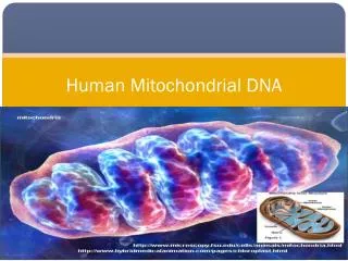
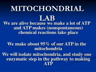
MITOCHONDRIAL LAB. We are alive because we make a lot of ATP and ATP makes (nonspontaneous) chemical reactions take place We make about 95% of our ATP in the mitochondria We will isolate mitochondria, and study one enzymatic step in the pathway to making ATP. Mitochondria and disease :.

E N D
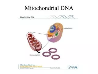
MITOCHONDRIAL LAB We are alive because we make a lot of ATP and ATP makes (nonspontaneous) chemical reactions take place We make about 95% of our ATP in the mitochondria We will isolate mitochondria, and study one enzymatic step in the pathway to making ATP
Mitochondria and disease: • CU MED CENTER: MITOCHONDRIAL MALFUNCTION LEADS TO PARKINSON’S DISEASE (SHAKING AS YOU AGE) • IF YOU RESTRICT YOUR DIET, YOU MAY LIVER LONGER & HEALTHIER; MORE CALORIES TAKEN IN, MORE NAD+ IS USED TO CARRY ELECTRONS (NOTE THAT FAD ALSO CARRIES ELECTRONS). HOWEVER, NAD+ IS ALSO NEEDED FOR THE SIR2 PROTEIN THAT PREVENTS AGING SYMPTOMS. THUS, LOTS OF SUGAR INTAKE, NAD+ IS NOT AVAILABLE TO SIR2– YOU AGE FASTER. • Bad mitochondria may be related to Diabetes
How we make most of our ATP: AEROBIC RESPIRATION: GLUCOSE IS BROKEN DOWN (HIGH ENERGY CHEMICAL BONDS BROKEN, ATOMS REMOVED) TO STRIP OFF ENERGETIC ELECTRONS.ENERGY FROM ELECTRONS IS USED TO MAKE ATP in the MITOCHONDRIAOXYGEN (02) ACCEPTS THE SPENT (LOW ENERGY) ELECTRONS= AEROBIC
PARTS OF AEROBIC RESPIRATION: • GLYCOLYSIS occurs in the cytoplasm (glucose broken in HALF to produce 2 pyruvate molecules) (ch. 9) (some list other “step” as ‘intermediate step”; moving pyruvic acid into the mitochondrion) • TCA CYCLE (or Kreb’s cycle)- where what is left of glucose is broken all the way down to C02 and all the electrons are stripped off • Electrons are carried (by NADH or FADH2) to the electron transport chain and ATP synthase where ATP is made from electron energy (ch. 10)
PARTS OF AEROBIC RESPIRATION Part 1. 2 Part 3
CH. 9: GLYCOLYSIS • GLYCOLYSIS IS A SERIES OF 12 CHEMICAL REACTIONS OCCURING IN THE CYTOPLASM • TAKE GLUCOSE (LIKE JET FUEL) AND STRIPS OFF ITS ELECTRONS IN THE HIGH ENERGY COVALENT BONDS • THIS BREAKS THE COVALENT BONDS AND BREAKS GLUCOSE IN HALF PRODUCING TWO 3 CARBON MOLECULES CALLED PYRUVATE • LATER, ENERGY FROM THESE ELECTRONS WILL BE USED TO MAKE ATP
ENERGETIC ELECTRONS TAKEN FROM GLUCOSEGIVEN TO NAD+WHICH CARRIES ELECTRONS INTO MITOCHONDRIA ELECTRONS GIVEN TO NAD is a REDUCTIONUSE MNEMONIC: OILRIG
Ch. 10: Last 2 Parts of Aerobic Respiration take place in the Mitochondrion. ATP made at inner membrane FOLDING MEMBRANE DOES WHAT? (HINT: GUT FOLDS)
Last 2 Parts involve the Electron Transport Chain (where electrons are stripped of their energy, energy used to pump Protons H+)Followed by allowing H+ to move back, turning ATP Synthase to make ATP. See SUMMARY ANIMATION FROM OUR TEXTBOOK…D:\cell biol 3611\mito respiration\respiration 1418m.mov
Electron transport animation from “Virtual cell” web site –see LINK ON our Cell Lab web siteLAST STEP: ATP SYNTHASE IS LIKE A LITTLE MOLECULAR TURBINE; TURBINE IS ROTATED BY MOVEMENT OF H+; THEN TURBINE MAKES ATP. Animations: • D:\cell biol 3611\mito respiration\ETCAdvanced.wmv
ANIMATIONS OF CHEMIOSMOSIS • D:\cell biol 3611\mito respiration\chemiosmosis2.swf • D:\cell biol 3611\mito respiration\ATP SYNTHASE MBC 14_1.mov • D:\cell biol 3611\mito respiration\17 ELEC TRANS CHAIN.MPG • D:\cell biol 3611\mito respiration\ATPGradientAdvanced.wmv (NOTE THAT SOME ANIMATIONS TALK ONLY OF THE H+ CONCENTRATION GRADIENT, IGNORING THE VERY IMPORTANT ELECTRICAL GRADIENT FOR THE H+!!)
We will study one enzyme in the TCA Cycle (see Ch. 10; esp figures used here) Succinate Dehydrogenasethis enzyme breaks two chemical bonds and removes two H atoms from what is left of glucose.
Succinate Dehydrog. Is located here… Succin Dehydrog Actually binds Membrane proteins Of the Electron Transport Chain
Pyruvate comes in from the cytoplasm into the mitochondrion, it is broken down in the TCA cycle to C02 and water. We will study TCA step 6…
In this reaction, once again what is left of glucose is broken down further by breaking bonds and removal of 2 H atoms. FADH2 carries the excited electrons to the electron transport chain (to make ATP from electron energy)
Better view of reaction… note the two H atoms that are removed are on different carbons and on opposite sides (trans, not cis) In the lab, the electrons are not given to FAD, but we add a dye that changes its absorbance when it takes the electrons (change in absorbance recorded by spectrophotometer)
Succinate Dehydrogenase is an enzyme; substrate succinate binds in the active site (similar to enzymes below)
Characteristics of Succ. Dehyd. • As it breaks chemical bonds between Carbon and Hydrogen (C-H) in succinate, it takes the excited electrons and the Hydrogen atoms (actually hydride) from the chemical bonds and gives them to FAD • FAD becomes FADH2 • FADH2 transfers the electrons to the electron transport chain. • Energy from excited electrons used to make ATP
Cont’d • Succ. Dehyd. Is an Integral (?) membrane protein in the inner mitochondrial membrane (hard to remove from membrane, hard to study) • All other TCA cycle enzymes are soluble (located in the matrix) • If we add a “reducible dye,” the dye not FAD will pick up the electrons • OILRIG: oxidation is loss of electrons, reduction is gain of electrons. • So, dye (or FAD) is reduced, succinate is oxidized to fumarate
Cont’d • Succinate dehyd. has a size of 100,000 daltons. Average protein is 50,000 daltons ; how many amino acids in succ dehy? (/100) • Also contains 8 iron atoms, • Fe (iron) atoms help in the transfer of electrons from succinate to FAD. • Has two subunits (so it has quarternary structure) • Has higher activity than any other TCA cycle enzyme
Succinate Dehydrogenase is turned on or off through allosteric regulation (page 144-145)…. • Allosteric regulation is how the body controls an enzyme (competitive inhibition is typically artificial or external to the body) • ATP or reduced coenzyme Q are allosteric activators of Succ Dehyd • Allosteric activators typically bind somewhere between the subunits of Succ Dehyd (not the active site) to stimulate the enzyme activity • Allosteric inhibitors act similarly to inhibit
Competitive Inhibition • Inhibitor resembles Substrate • This is not how the body/cell regulates enzymes (typically) - some medicines work this way • So this method is “artificial” and used in test tubes to study an enzyme • The inhibitor can bind to the active site (preventing the normal substrate from binding) but the inhibitor cannot form the product • So, both the inhibitor and Substrate competefor the active site of the enzyme • If the substrate is in excess, the inhibitor will not inhibit
Competitive Inhibitors resemble the normal substrate (but cannot be turned into product—so they tie up enzymes by binding to their active site) Malonate
Competitive Inhibitors resemble the normal substrate -but cannot be turned into product—so they tie up enzymes by binding to their active site.Malonate is a molecule that looks like succinate, but it cannot be made into fumaric acid (product) so malonate is a competitive inhibitor. Malonate is in a COMPETITION for the active site of the enzyme with succinate-- which ever is in higher concentration typically wins!Some medicines are competitive inhibitors
Other competitive inhibitors… • Other “dibasic acids” (means that they have two carboxylic acid functional groups= C00-) can act as competitive inhibitors • The other dibasic acids inhibit because the distance between the two C00- is about the same as the distance in succinate. • The active site of succinate dehydrogenase must have two + charges that are separated by the same distance
Note that as long as the spacing between the two – ends is ~same as in Succinate, get competitive inhibition. Even two negative charges of pyrophosphate can act as a negative inhibitor:
SUCCINATE FITS INTO ACTIVE SITE (SOME OTHER “DIBASIC ACIDS” HAVE SAME SPACING BETWEEN NEGATIVE CHARGES) 0 = C - C - C – C = 0 0- 0- + + ACTIVE SITE- WHERE SUBSTRATE OR COMPETITIVE INHIBITORS BIND. HERE, FIND AMINO ACIDS WHERE THEIR R GROUP HAS + CHARGE SUCCINATE DEHYDROGENASE
So, we will isolate mitochondria using centrifugation, and study Succinate DehydrogenaseTo Isolate Organelles, you homogenize the cell and then use centrifugtation to Isolate the organelle
A rotor moves round and round, and heavy particles move to the bottom of the test tube fasterText: p. 322-323, Sixth Ed Differential Centrifugation
So, we will 1. isolate mitochondria from Xenopus liver and 2. follow Succinate Dehydrogenase activity by adding Succinate 3. add a competitive inhibitor called Malonate to reduce Succ Dehyd activity
Enzyme Kinetics • If little substrate is around, there will be very little enzyme activity and the rate of the reaction will be slow • If there is more substrate around, the enzyme will be more active and the reaction will be faster • At a certain point, even if you raise the substrate concentration further, the rate of the reaction will not increase • THIS IS SATURATION KINETICS (page 136 to 139 in text; 6th ed)
Rate of Reaction (slope of OD600 vs. time) Saturation at higher substrate concentrations because all enzyme working as hard as they can- the enzymes Are saturated!
Vm = maximum velocity or rate of the reactionKm = a measure of enzyme-substrate affinity (low Km means high affinity) • Obtain Vm by going over from the Y axis (rate of the reaction when it first begins) to where the “rectangular hyperbola” levels off • Obtain Km by going down the Y axis to one half Vm, then going over to the line in the graph, then going down to X axis. • You can also obtain the values by using a Double Reciprocal Plot (Fig. 6-12 and 6-13).
= [Succinate] Rate is initial slope for each concentration of succinate (in OD600 Versus time)
Double Reciprocal Plot