Understanding Anemia: Diagnosis and Management in Clinical Practice
Anemia is characterized by a reduction in hemoglobin or red blood cell volume below normal limits, influenced by factors such as age and gender. Causes include decreased production and increased loss. A thorough history and physical examination are essential, looking for signs of blood loss, family history, and dietary factors. Laboratory studies, including hemoglobin, hematocrit, and reticulocyte counts, are critical in diagnosing different anemias (microcytic, normocytic, etc.). This guide outlines the approach to diagnosis and the interpretation of laboratory results.

Understanding Anemia: Diagnosis and Management in Clinical Practice
E N D
Presentation Transcript
Anemia Paolo Aquino PGY-I January 2005 VA Hospital
Definition • Anemia is a reduction of hemoglobin or volume of red blood cells from normal limits • Variation due to age, gender • Decreased production • Increased loss
History • Signs of blood loss • Onset- rapid vs. gradual • Family history • Exposures • Diet • History of infection • PMH- cancer, renal, endocrine
Physical exam • Appearance • Vital signs • HEENT • Heart • Abdominal
Laboratory studies • Hemoglobin and hematocrit • Mean corpuscular volume • Reticulocytes • Peripheral smear
Microcytic anemia • Most common cause: iron-deficiency anemia • Thalassemia • Lead poisoning • Sideroblastic anemia • Anemia of chronic disease
Normocytic anemia • Marrow hypoplasia • Marrow infiltration • Myelofibrosis • Renal insufficiency • Anemia of chronic disease • Mild iron deficiency • Mixed microcytic/macrocytic
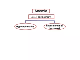
Approach • History and physical • Hemoglobin, hematocrit • Reticulocytes decreased production, increased loss • Remember to correct the retic count for the anemia
Approach • If reticulocytes low, check MCV • Low MCV: check iron studies, examine peripheral blood smear, hemoglobin electrophoresis • Normal MCV: check iron studies, examine peripheral blood smear, test endocrine function, consider bone marrow aspiration
Peripheral smear • Normal RBC size equal to nucleus of a mature lymphocyte • Shape • Spherocytes • Sickle cell • Helmet cells • Tear drop cells
Peripheral smear • Color • Hypochromasia • Iron deficiency • Sideroblastic • Hyperchromasia • Megaloblastic anemia • Spherocytosis
Peripheral smear • Morphology • Howell-Jolly bodies • Basophilic stippling • Heinz bodies • Requires supravital stain • Cabot ring • Nuclear remnants
Peripheral smear • Morphology • Rouleaux formation • Parasites • Nucleated RBCs • Target cells
Summary • Once anemia is established • Check reticulocyte count • Correct count for anemia • Increased retic count indicates RBC loss- hemolysis or bleed • Decreased retic count indicates decreased RBC production • Check MCV
Summary • Decreased reticulocyte count • Normocytic • check iron studies, examine peripheral blood smear, test endocrine function, consider bone marrow aspiration • Microcytic • check iron studies, examine peripheral blood smear, hemoglobin electrophoresis