Download
1 / 61
700 likes | 1.17k Vues
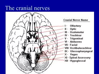
Anatomy of the Eye & the 12 cranial nerves. 12 Cranial Nerves. I Olfactory II Optic III Oculomotor IV Trochlear V Trigeminal VI Abducens VII Facial VIII Auditory ( Vestiblochlear ) IX Glossopharyngeal X Vagus XI Accessory XII Hypoglossal.

E N D
12 Cranial Nerves I Olfactory II Optic III Oculomotor IV Trochlear V Trigeminal VI Abducens VII Facial VIII Auditory (Vestiblochlear) IX Glossopharyngeal X Vagus XI Accessory XII Hypoglossal
There are 12 pairs of cranial nerves. I Olfactory II Optic III Oculomotor IV Trochlear V Trigeminal VI Abducens VII Facial VIII Auditory (Vestiblochlear) IX Glossopharyngeal X VagusXI Accessory XII Hypoglossal
Anatomy of the Eye Characteristics • Measures about 1” and is shaped as a sphere • See only anterior 1/6 Accessory Structures • Extrinsic Eye Muscles • Eyelids • Conjunctiva • Lacrimal apparatus
Eyebrow Site where conjunctiva merges with cornea Eyelid Eyelashes Pupil Palpebral fissure Lacrimal caruncle Medial commissure (canthus) Lateral commissure (canthus) Sclera (covered by conjunctiva) Iris Eyelid Figure 8.1
Anatomy of the Eye Anterior Aspects of the Eye • Eyelids- provide protection • Medial/Lateral Commissure (corners of the Eye) • Palpebral fissure- space b/w eyelids • Eyelash- extends from the eyelid • Tarsal glands- Modified sebacious gland • Produces oily secretion - lubrication • Ciliary glands – modified sweat gland • Found between the eyelashes • Conjunctive – lines eyelid and covers the outer surface of the eyeball • Secretes mucous
Excretory duct of lacrimal gland Lacrimal gland Conjunctiva Anterior aspect Eyelid Eyelashes Tarsal glands Eyelid Figure 8.2a
Homeostatic Imbalance of the Eye Characteristics • Inflammation of the conjunctiva • Infectious conjunctivitis Conjunctivitis Pink Eye
Lacrimal sac Lacrimal gland Excretory ducts of lacrimal gland Lacrimal canaliculus Nasolacrimal duct Inferior meatus of nasal cavity Nostril (b) Figure 8.2b
Lacrimal Apparatus 1. Consists of the lacrimal glands and ducts to drain the secretions into the nasal cavity 2. Lacrimal Glands • Located above the lateral end of each eye • Releases tears- dilute salt solution • Tears flush the eye across the canaliculi medially into the LacrimalCanaliculi • LacrimalCanaliculi a. Sends tears into the Lacrimal sac b. Lacrimal sac receives the tears • Nasolacrimal Duct a. empties the tears into the Nasal Cavity
Lacrimal Secretions 5. Lacrimal Secretions (Tears) contain mucuos, Antibodies and Lysozyme (destroy bacteria) 6. Nasal Mucosa • Connects with the lacrimal duct system • Effects of Nasal Mucosa will reach the eye
6 Extrinsic Eye Muscles 1. Attach to the outer surface of the eye 2. Produce Gross eye movment
Superioroblique muscle Superioroblique tendon Superiorrectus muscle Conjunctiva Lateral rectusmuscle Inferiorrectusmuscle Inferiorobliquemuscle Opticnerve (a) Figure 8.3a
Trochlea Superioroblique muscle Superioroblique tendon Axis at center of eye Superiorrectus muscle Inferior rectus muscle Medialrectus muscle Lateralrectus muscle (b) Figure 8.3b
Eyeball 1. Hollow Sphere • 2. Composed of 3 layers • a. Fibrous layer - Outside layer • b. Vascular layer - Middle layer • c. Sensory layer - Inside layer 3. Inner sphere filled with fluid called Humors 4. Lens – supported upright within the cavity • a. Divides the eye into 2 chambers • 1. Aqueous humor – Anterior Chamber • 2. Vitreous Humor – Posterior chamber
Layers forming the eye wall • 1. Fibrous Layer – outermost layer • A. Sclera – protective layer • 1. Thick glistening white connective tissue • 2. seen in the anterior as the “white of the eye” • B. Cornea • 1. the central anterior portion of the fibrous layer • 2. crystal clear (window of the eye) – light enters • 3. many nerve endings – pain fibers • 4. If touched, blinking and tearing occur • 5. Exposed part of the eye • a. subject to injury • b. great ability to repair • 6. only body tissue that can be transplanted without rejection • a. no blood supply – no immune system
Layers forming the eye wall • 2. Vascular Layer (Choroid)– middle layer of the eyeball • a. Blood rich nutritive tonic that contains dark pigment • b. Prevents the scattering of light inside the eye • c. Anteriorly, modified to form two smooth muscle • structures • 1. Ciliary body – attaches to lens by ligamentscalled • CiliaryZonule 2. Iris – filled with pigment (circular and radial smooth muscle) 3. Pupil – rounded opening to the ris a. Bright light and Close vision 1. Pupils constrict (contraction of circular muscles) b. Dark and far vision 1. Pupils enlarge (radial fibers contract)
Layers forming the eye wall • 3. Sensory Layer (Retina) • Retina – innermost 2 layered retina • a. Extends anterior to the ciliated body • b. Outer Layer – pigmented • 1. Prevents the scattering of light inside • the eye like the choroid • 2. Acts as Phagocytes • a. remove dead/ damaged receptor • cells • b. stores Vitamin A • c.Inner Layer – Neural layer - transparent
Sensory Layer (Continued) • c.Inner Layer – Neural layer – transparent • 1.contains millions of receptor Cells • called Photoreceptors(Rods and Cones) • 2. Impulse pathway travels from: • (2 neuron chain) • photoreceptors bipolar cells • ganglion cellsOptic Nerve • 3. Leave the retina via the optic nerve and travel to the optic cortex
Sclera Ciliary body Choroid Ciliary zonule Retina Cornea Fovea centralis Iris Pupil Optic nerve Aqueous humor (inanterior segment) Lens Scleral venous sinus (canal of Schlemm) Central artery and vein of the retina Vitreous humor (in posterior segment) Optic disc (blind spot) (a) Figure 8.4a
Ciliary body Vitreous humor in posterior segment Iris Retina Margin of pupil Choroid Sclera Aqueous humor (in anteriorsegment) Fovea centralis Optic disc Optic nerve Lens Cornea Ciliary zonule (b) Figure 8.4b
Figure 8.4internal anatomy of the eye (sagittal section). Sclera Ciliary body Choroid Ciliary zonule Retina Cornea Fovea centralis Iris Pupil Optic nerve Aqueous humor (in anterior segment) Lens Scleral venous sinus (canal of Schlemm) Central artery and vein of the retina Vitreous humor (in posterior segment) Optic disc (blind spot) (a) Ciliary body Vitreous humor in posterior segment Iris Retina Margin of pupil Choroid Sclera Aqueous humor (in anterior segment) Fovea centralis Optic disc Optic nerve Lens Cornea Ciliary zonule (b)
Photoreceptor cells (rods & cones) • 1. Distributed over the entire Retina except where the optic • nerve leaves the eye • 2. Optic disc – (the point where the optic nerve leaves the eye) • a. Blind spot • 3. Rods and Cones are not evenly distributed • a. Rods – dense at the peripheral edge of the retina • decrease in number as you move to the center of • the Retina • 1. see shades of gray in dim light • 2. allows for peripheral vision • 3. night blindness – results from an • interference with rod function • Causes • a. Vit A deficiency • b. Leads to a deterioration of the • neural retina tissue • c. Vit A can help to restore function if taken • PRIOR to degeneration
Photoreceptor cells (cont’d) • 3. (Continued) • b. Cones • 1. allows us to see details and colors in • bright light • 2. most dense in the center of the Retina • 4. Fovea Centralis • a. Lateral to each blind spot • b. tiny pit containing only Cones • c. area of greatest visual acuity • (sharpness)
Figure 8.5a The three major types of neurons composing the retina. Pigmented layer of retina Neural Layer Rod Cone 2 ganlion chain Bipolar cells Pathwayof light Ganglion cells (a)
Rhodopsin (purple pigment in Rods) a. formed from union of Opsin and Retinal (modified Vit A) Kinked shape. b. Retinal straightens when hit with light (purple color changes to yellow (bleaching) c. Once colorless, the Retinal is now Vit A again d. once the Vit A returns to its kinked form, it combines with Opsin to regenerate into Rhodopsin (An ATP-requiring process) Light Light Light A Closer Look 8.1 Visual Pigments—the Actual Photoreceptors Process of bipolar cell Synaptic endings Inner fibers Rod cell body Rod cell body Cone cell body Outer segment – attached to the cell body, light trapping contains visual discs to trap light. a. bleaching – results from stimulation of light, pigment regenerates b. this causes electrical changes in the photoreceptor cells-nerve impulse sent to brain Nuclei Outer fiber Mitochondria Inner segment Retinal (visual yellow) Discs containing visual pigments Pigmented layer Light absorption causes Releases Outer segment Opsin Rhodopsin (visual purple) Pigment cell nucleus Melanin granules Bleaching of the pigment
Figure 8.5b The three major types of neurons composing the retina. Pigmentedlayer of retina Neural layerof retina Centralarteryand veinof retina Opticdisc Sclera Opticnerve Choroid (b)
5. Macula (macula lutea) (from Latin macula, "spot" + lutea, “yellow") a. is an oval-shaped highly pigmented yellow spot near the center of the retina b. It has a diameter of around 1.5 mm c. defined as having two or more layers of ganglion cells d. Fovea Centralis is located near the center a. contains the largest concentration of cone cells in the eye and is responsible for central, high resolution vision.
e. Because the macula is yellow in color it absorbs excess blue and ultraviolet light that enter the eye 1. acts as a natural sunblock (analogous to sunglasses) f. The yellow color comes from its content of lutein and Zeaxanthin a. Zeaxanthin is found mostly at the macula b. Lutein found in the retina. c. There is some evidence that these carotenoids protect the pigmented region from some types of macula degeneration
Macula Bloodvessels Retina Optic disc Foveacentralis Figure 8.8 The posterior wall (fundus) of the retina as seen with an ophthalmoscope. Lateral Medial
Macular Degeneration 1. There is a loss of peripheral vision 2. it may go unnoticed for some time 3. damage will result in loss of central vision
3 Types of cones • Each most sensitive to a particular wavelength of light • 1. Blue • 2. Green • 3. Green and Red - called the red cones, only respond • to red • Intermediate colors • Multiple impulses yield a blend of colors as interpreted by the visual corex • Blue/Red Purple • when all 3 cones are stimulated eyes • will result in White color • color mix occurs at the Brain Red • Yellow • Green
Figure 8.6 Sensitivities of the 3 cone types to the different wavelengths of visible light. 560 nm (red cones) 530 nm (green cones) 420 nm (blue cones) Light absorption by cone populations 450 500 550 600 650 700 750 380 Wavelengths (nanometers)
Lens • 1. focuses light to the Retina • 2. Biconvex crystal like structure • 3. Held upright in position by suspensory ligaments called • (CiliaryZonules) which attach to the Ciliary body. • Lens Divides the eye into 2 chambers: • 1. Anterior (Aqueous) segment • a. contains clear fluid called Aqueous Humor • b. Reabsorbed into the venous blood through the • Sclera Venous sinus or (Canal of Schlemm) • 1. located at the junction of the Sclera/Cornea • 2. Posterior (Vitreous) segment • 1. Contains clear fluid called Vitreous Humor (Body) • 2. Prevents collapse of the eyeball • 3. Maintains intraocular pressure • 4. Provides nutients for lens/cornea
Lens • Imbalance Disorders • Color blindness • 1. The lack of all 3 cones • 2. Most common – lack of red or green receptors • a. 2 colors seen as one, depends on the cone • b. gene for color vision on X chromosome • c. sex linked – seen more in males • Cataracts • 1. Hard opaque hazy distorted appearance • 2. results in blindness • 3. Risk factors • a. Diabetes, sunlight, smoking • 4. treatment – surgery, lens replacement • Glaucoma • 1. results from an increase of the pressure in the eye • 2. due to a build up of Aqueous Humor • 3. Test: Tonometer (puff of air)- measures the internal pressure of the eye
Ophthalmoscopic exam • Ophthalmoscope • Instrument that illuminates the interior of the eye • Able to view the Retina, Optic disc, Blood vessels at the Fundus, Macula, Fovea Centralis • Fundus Exam – used to detect pathology • Diabetes – vascular blotches (micro aneurisms) • Arteiosclerosis – copper wiring reflex (Hypertensive Retina) • Degeneration of the optic nerve and retina • See pale optic nerve – loss of axons & myelin
Diabetic Retinopathy • Seemicroanneurisms (blotches) • Hard exudates (yellow) and cotton wool spots (white) • Macular Degeneration – pigmented spotting on Retina
Macula Bloodvessels Retina Optic disc Foveacentralis Figure 8.8 The posterior wall (fundus) of the retina as seen with an ophthalmoscope. Lateral Medial
Fundus photographs of the right eye (left image) and left eye (right image). The gaze is into the camera, so in each picture the macula is in the center of the image, and the optic disk is located towards the nose.
Hypertensive retinopahy AV nicking
Copper wire reflex AV nicking Hypertensive retinopathy with AV nicking and mild vascular tortuosity
physiology of vision • Distant Objects • Set for distant vision • Light from over 20 feet away approaches as parallel rays • Lens does not need to change shape for focus • Closer Objects • Light tends to scatter, diverge – spread out • Lens must bulge more • A. Ciliary body contracts • B. Allows lens to become more convex Resting Eye