Download
1 / 58
590 likes | 879 Vues
HEMATOPOIETIC STEM CELL TRANSPLANTATION (HSCT) IMED 7190 - Medical Immunology Cindy Ellison, PhD, D(ABHI) Departments of Pathology and Immunology. Objectives: Review HLA basics: structure, function, nomenclature and typing methods.

E N D
HEMATOPOIETIC STEM CELL TRANSPLANTATION (HSCT) IMED 7190 - Medical Immunology Cindy Ellison, PhD, D(ABHI) Departments of Pathology and Immunology
Objectives: • Review HLA basics: structure, function, nomenclature and typing methods. • 2. Review the role of HSCT in the treatment of hematological malignancies and other diseases. • 3. Understand the potential benefits and complications of allogeneic HSCT. • 4. Understand the immunobiology of graft-versus-host disease (GVHD) and the graft-versus-malignancy effect. • 5. Review strategies for preventing the development of GVHD in patients receiving an allogeneic HSCT.
Major Histocompatibility Complex (MHC) • multigenic system, the most gene-dense region in our genome • highly variable between individuals, multiple alleles exist • proteins encoded by the MHC are known as the human leukocyte antigens (HLA) • located on chromosome 6
Organization of HLA genes on chromosome 6 DP DQ DR B C A Complement TNF LT Class III Cytokines Class I Class II Centromere Telomere
Structure of HLA proteins CLASS II CLASS I peptide peptide H2N NH2 1 domain 2 domain NH2 a chain b chain a chain NH2 2 microglobulin 3 domain HOOC COOH HOOC COOH
Classical MHC genes • genes encoding proteins involved in the presentation of antigen to T cells • Class I MHC • molecules present on virtually all nucleated cells • Humans: HLA-A, HLA-B and HLA-C
Classical MHC genes • Class II MHC • molecules expressed on “antigen presenting cells” (APC) of the immune system • e.g. macrophages, B cells and dendritic cells • Humans: HLA-DR, HLA-DQ and HLA-DP
Other Class I Molecules • Collectively, these are referred to as “non-classical” class I genes or “class I-related genes”. • HLA-E, -F, -G and –H • less polymorphic, often invariant
MHC diagram,viewed from above • Many of the amino acid substitutions contributing to polymorphism appear on the -pleated sheet or “floor” of the groove, and on the inner surfaces of the helices, i.e., the places where the MHC molecule contacts antigenic peptide.
Mutations • MHC genes are a “hot spot” for mutations. The frequency is twice that seen at non-MHC loci. This results in increased diversity in the MHC genes. • New alleles are discovered nearly every day. Some of these result in amino acid substitutions and some do not. Some are located in the exons and some are in the introns. These factors are reflected in the name of the new allele.
Inheritance of MHC Genes • Genes of the MHC are co-dominantly expressed on lymphocytes of the progeny. For example, you express one HLA-A gene from your mother and one from your father on a nucleated cell in your body. Haplotype • the collection of HLA genes from one chromosome • Certain alleles and haplotypes can be present at higher frequencies in populations that are isolated geographically or in small ethnic groups.
Family Haplotypes Possible combinations in offspring Dad Mom a b c d a c a d b c b d Patient’s HLA typing reveals: b d For each sibling there is a 25% chance that he or she will be HLA-identical with the patient.
Example HLA serological typing for class I reveals the following HLA types: Patient: A2, A11; B7, B35; Cw7, Cw4 Mother: A2, A29; B7, B44; Cw7, Cw16 Father: A11, A24; B35, B7; Cw4, Cw7 What two haplotypes does the patient have?
Haplotype 1: A2, B7, Cw7 Haplotype 2: A11, B35, Cw4
Recombination between homologous chromosomes A A A A A A C C C C C C B B B B B B DRB1 DRB1 DRB1 DRB1 DRB1 DRB1 DQB1 DQB1 DQB1 DQB1 DQB1 DQB1 • Frequency of recombination across the MHC (HLA-DPB1-HLA-A) is 2-2.5%. Recombination is not seen between HLA-B and C or between HLA-DQA and DRB1. (Reviewed by M. Carrington in Immunological Reviews, 167:245; 1999). .
Why is HLA matching important? …it is the best way to prevent the development of graft-versus-host disease, a serious and potentially fatal complication of allogeneic HSCT
Clinically significant GVHD develops in about 40% or HLA-matched sibling transplants and in approximately 80% of all HLA-matched unrelated donor transplant recipients.
Serological vs. Molecular Typing • Serological typing: • Lymphocytes are incubated with anti-HLA antibodies (antisera from sensitized individuals), followed by complement. Cells recognized by the antibody are killed by complement-mediated lysis (microlymphocytotoxicity assay). If >90% of the cells in a well are lysed, it is assumed that the cells in that particular well carried the HLA molecule recognized by the typing antibody. • Typing is reported based on the antigens identified by the antibodies, e.g. A2, A11. Some antibodies recognize groups of antigens, e.g. B40 (B60, B61).
Molecular Typing • Genomic DNA is extracted from blood and amplified using the polymerase chain reaction (PCR) ds DNA 3’ 5’ 5’ 3’ 3’ 5’ Denaturation P2 P1 3’ 5’ 5’ 3’ Annealing 3’ 5’ 5’ 3’ Extension 5’ 3’
Molecular Typing • HLA antigens and alleles are identified using a number of different methods. The level of resolution depends on the method(s) used. • SSP: specific primers bind to polymorphic regions • SSO: specific probes bind to polymorphic regions • SBT: nucleic acid sequence is determined
Primers vs. Probes Both are short single stranded pieces of DNA that bind to specific, complementary DNA sequences. Primers are used for synthesizing DNA Probes bind to complementary sequences of DNA and permit detection by labeling
Sequence Specific Priming (SSP) Sequence-specific primers bind to ss DNA Sequence-specific primers do not bind to ss DNA Amplification by PCR (Positive Result) No amplification by PCR (Negative Result)
Reverse Sequence Specific Oligonucleotide Probing Method Substrate Enzyme SA biotin Amplified DNA Probe Membrane
HLA Nomenclature HLA-A*02:01 Specific HLA locus Indicates HLA region and serves as a prefix for the HLA gene A group of alleles which encode the A2 antigen A specific HLA allele Suffixes Null alleles: alleles that encode proteins that are not expressed, e.g., HLA-A*2409N (can be confirmed by serological typing) Low expression alleles: alleles that encode a protein with reduced surface expression, e.g. A*3014L
Finding A Suitable Donor Lower risk of complications with autologous transplants when compared to allogeneic transplants, but there is a greater risk of relapse. • An HLA-matched sibling is still regarded as the ideal donor but a matched, unrelated donor (MUD) can be used if an HLA-matched sibling is unavailable. This is currently the most effective way to prevent graft-versus-host disease. Comparable results are being seen with appropriately matched umbilical cord blood units. HLA matching requirements are less stringent since cord blood-derived products have a diminished ability to induce GVHD. • Odds of a sibling matching the patient are 25%.
In Manitoba, donor and recipient are typed for HLA-A, -B, C, DRB1 and DQB1 with the aim of finding a 10/10 allele-level match. If no suitable HLA-matched sibling donor is found, a search is initiated for an HLA-matched donor in the unrelated registries. • An unrelated HLA-matched donor can be identified for 80% of Caucasian patients but the percentage is lower for other ethnic groups.
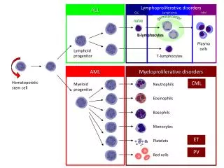
Sources of Hematopoietic Stem Cells 1. Bone marrow Leger, CS and Nevill, TJ CMAJ 2004; 1569-1577
2. Peripheral Blood Collect Peripheral Blood Progenitor Cells (PBPC) by leukapheresis Granulocyte Colony Stimulating Factor (G-CSF) 5-10 g/kg on day -5 to day -1
Conditioning Regimen High-dose chemotherapy and sometimes radiotherapy is given to eradicate malignant cells and/or to suppress the patient’s ability to reject the donor’s stem cell product. Conditioning is given over a one-week period. The patient becomes pancytopenic and develops other non-hematological side-effects collectively referred to as “regimen-related toxicities”.
Fig. 3: Oropharyngeal mucositis, which may occur as a result of conditioning before stem cell transplantation Leger, C. S. et al. CMAJ 2004;170:1569-1577
Reduced Intensity Conditioning: Can be used instead of myeloablative conditioning Less toxic Allows transplants to be performed on older patients and younger patients with significant co-morbidities
Infusion of the stem cell product Leger, CS and Nevill, TJ CMAJ 2004; 1569-1577
Engraftment • Donor stem cells home to the patient’s bone marrow cavity and begin to differentiate into the various blood cell lineages. • Primary graft failure or graft rejection are serious complications that occur in less than 5% of recipients (less often with HLA-matched sibling transplants). • Immune reconstitution may take 12 months or longer. As it occurs, the risk of opportunistic infection decreases. As the numbers of donor T cells increase, so does the risk of graft-versus-host disease (GVHD).
Graft-versus-Host Disease • Conditions for a GVH Reaction: • The graft must be immunocompetent. • The recipient must be unable to reject the graft. • The donor and recipient must be genetically non-identical, particularly with respect to major histocompatibility antigens.
Clinical Course of GVHD Acute • epithelial lesions in skin, intestine, liver, lung and bile duct • develops before day 100 post-transplantation • mediated primarily by Th1 cytokines (in mice) Chronic • sicca/Sjogrens-like syndrome, scelroderma-like lesions • develops after day 100 post-transplantation • mediated primarily by Th2 cytokines (in mice)
Acute cutaneous GVHD Leger, CS and Nevill, TJ CMAJ 2004; 1569-1577
Donor T cells recognize allogeneic HLA in the patient (if HLA mismatches exist). Minor histocompatibility antigens may also be involved because GVHD still develops in patients receiving a transplant from an HLA-matched sibling. • Approximately 10% of all T cells in an individual react with alloantigen, but less than 1% of all T cells in an individual react with any single antigenic peptide. • T cell receptors recognize specific allogeneic class I and II HLA molecules +/- peptide • Donor CD8+ cytotoxic T cells contribute to the graft-versus-malignancy effect
The development of GVHD can be broken down into 5 basic steps. (Reviewed by Socie and Blazar, Blood 114:4327, 2009)
ICOSL OX40L CD80/ CD86 CD80/ CD86 4-1BBL CD153 CD40 4-1BB CD30 CD28 ICOS CD40L OX40 CTLA4
Pathogenesis of Acute GVHD Allogeneic Response Cellular and cytokine mediators Tissue Injury GVHD Endotoxin (LPS)
B A Role of intestinal epithelial cell apoptosis and LPS
Non-HLA Polymorphisms • The following have been implicated as risk factors for the development of GVHD in some studies: • cytokine polymorphisms (e.g. TNFa, IL-10 and IFNg) • TLR polymorphisms • KIR expression--Donor NK cells receive inhibitory and activatory cells when they engage HLA-C and other KIR ligands on the patients cells. In some studies, this has been shown to mediate GVM effects.