Download
1 / 44
440 likes | 607 Vues
Special-topic Lecture Bioinformatics: Modeling Cell Fate. Leistungspunkte/Credit points: 5 (V2/Ü1) This course is taught in English language. The material (from books and original literature) are provided online at the course website:

E N D
Special-topic Lecture Bioinformatics: Modeling Cell Fate Leistungspunkte/Credit points: 5 (V2/Ü1) This course is taught in English language. The material (from books and original literature) are provided online at the course website: http://gepard.bioinformatik.uni-saarland.de/teaching/ss-2013/stl-bioinformatics-modcellfate-ss13 Biological topics to be covered: This course will enter into details of three selected topics in current cell biology: (1) Cell cycle (2) Stem cell differentiation (3) Cancerogenesis Modeling of Cell Fate
Bioinformatics content • microarray expression analysis • DNA methylation analysis • GO and pathway annotation • interaction networks • application of clustering techniques • construction of gene-regulatory networks • stochastic simulations Modeling of Cell Fate
Aim of this lecture, „Lernziele“ (1) The aim of this course is not to fully cover these three topics but to enter deeply into various details of these fields. (2) This course should train you to analyze original biological data using modern bioinformatics tools. (3) You shoud also become familiar with the biological processes (pathways) controlling cellular adaptation / cell fate. Modeling of Cell Fate
Tutorial We will handout 6 biweekly assignments. Groups of up to two students can hand in a solved assignment. Send your solutions by e-mail to the responsible tutors : Mohamed Hamed, Ruslan Akulenko, Christian Spaniol until the time+date indicated on the assignment sheet. The tutorial on Thursday 12 am - 1 pm will provide help to understand the papers, prepare the student presentations and the assignment solutions. Schein condition 1 Only those students can get a „Schein“ who have obtained more than 50% of the points for all assignments. Modeling of Cell Fate
Schein = pass 3 written tests Schein condition 2 The successful participation in the lecture course („Schein“) will be certified upon fulfilling Schein condition 1 and upon successful completion of 3 written 45 minute tests. Each test roughly covers the content of one of the three lecture topics. Dates: probably at the beginning of lectures V5, V9, V13. All students registered for the course may participate in the tests. 2 out of 3 tests have to be passed. The final grade on the Schein is the average of your 2 best tests. Rounding scheme: (1.0 + 1.3 -> 1.0 ; 1.3 + 2.0 -> 1.7) Modeling of Cell Fate
written tests The tests will cover the lecture material (slides on the lecture website) and the theory behind the assignments for this topic. In case of illness please send E-mail to: kerstin.gronow-p@bioinformatik.uni-saarland.de and provide a medical certificate. Those who miss or fail one test, will be given a second-chance oral exam. If you fail or miss more than two tests, you cannot get a Schein. Modeling of Cell Fate
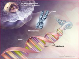
Gene Transcription etc. Basic terms that you should remember from an introductory genetics lecture ... or that you should read up: Genome Genes Introns, Exons Nucleus DNA-Polymerase Transcription mRNA Splicing Ribosome tRNA Translation Modeling of Cell Fate
A biological cell Schematic of typical animal cell, showing subcellular components. Organelles: (1) nucleolus (2) nucleus (3) ribosome (4) vesicle (5) rough endoplasmic reticulum (ER) (6) Golgi apparatus (7) Cytoskeleton (8) smooth ER (9) mitochondria (10) vacuole (11) cytoplasm (12) lysosome (13) centrioles HeLa cells stained for DNA with the Blue Hoechst dye. The central and rightmost cell are in interphase, thus their entire nuclei are labeled. On the left a cell is going through mitosis and its DNA has condensed ready for division. wikipedia.org Modeling of Cell Fate
cell cycle The cell cycle, or cell-division cycle, is the series of events that takes place in a cell leading to its division and duplication (replication). In cells without a nucleus (prokaryotes), the cell cycle occurs via a process termed binary fission. In cells with a nucleus (eukaryotes), the cell cycle can be divided in 2 brief periods: interphase—during which the cell grows, accumulating nutrients needed for mitosis and duplicating its DNA—and the mitosis (M) phase, during which the cell splits itself into two distinct cells, often called "daughter cells". Each turn of the cell cycle divides the chromosomes in a cell nucleus. www.wikipedia.org Modeling of Cell Fate
Phases • The cell cycle consists of 4 distinct phases: • - G1 phase, • - S phase (synthesis), • G2 phase • and M phase (mitosis). • Interphase: combines G1, S, and G2 • Activation of each phase is dependent on the proper progression and completion of the previous one. • Cells that have temporarily or reversibly stopped dividing are said to have entered a state of quiescence called G0 phase. Schematic of the cell cycle. Outer ring: I = Interphase, M = Mitosis; Inner ring: M = Mitosis, G1 = Gap 1, G2 = Gap 2, S = Synthesis. www.wikipedia.org Modeling of Cell Fate
Activity during 4 phases M phase itself is composed of 2 tightly coupled processes: - mitosis, in which the cell's chromosomes are divided between the two daughter cells, and - cytokinesis, in which the cell's cytoplasm divides in half forming distinct cells. www.wikipedia.org Modeling of Cell Fate
Regulation of the eukaryotic cell cycle Regulation of the cell cycle involves processes crucial to the survival of a cell, including the detection and repair of genetic damage as well as the prevention of uncontrolled cell division. The molecular events that control the cell cycle are ordered and directional. Each process occurs in a sequential fashion. It is impossible to "reverse" the cycle. Leland Hartwell Tim Hunt Paul Nurse Noble Price in Physiology/Medicine 2001 „for their discoveries of key regulators of the cell cycle“ Two key classes of regulatory molecules, cyclins and cyclin-dependent kinases (CDKs), determine a cell's progress through the cell cycle. www.wikipedia.org Modeling of Cell Fate
Cell cycle control model Tyson et al, Curr. Op. Cell Biol.15 (2003) 221 Modeling of Cell Fate 13
protein kinase A: a model system for phosphate transfer Susan S. Taylor UC San Diego Masterson et al. Nat Chem Biol. 6, 825 (2010) Taylor et al. Phil Trans R.Soc. B (1993) Modeling of Cell Fate 14
Cyclin – cdk2 complex crystal structure Cyclin A – cdk 2 complex red: PSTAIRE motif yellow: activation loop Nikola Pavletich Memorial Sloan-Kettering Cancer Center Cyclin A – cdk2 phosphorylated at Thr160 www.wikipedia.org Modeling of Cell Fate
Crystal structure p27 (Kip1) is shown bound to the CyclinA-Cdk2 complex, provoking profound changes in the kinase active site and rendering it inactive. p27 also interacts with the secondary substrate recognition site on the cyclin. p27(Kip1)-CyclinA-Cdk2 Complex www.wikipedia.org Modeling of Cell Fate
Cdk1-phosphorylation sites Cdk1 substrates frequently contain multiple phosphorylation sites that are clustered in regions of intrinsic disorder. Their exact position in the protein is often poorly conserved in evolution, indicating that precise positioning of phosphorylation is not required for regulation of the substrate. Cdk1 interacts with nine different cyclins throughout the cell cycle. Expression of human cyclins through the cell cycle. www.wikipedia.org Enserink and Kolodner Cell Division 2010 5:11 Modeling of Cell Fate www.wikipedia.org
The classical model of cell-cycle control Nature Reviews Molecular Cell Biology 9, 910-916 (2008) Cyclin-dependent kinases (cDKs) trigger the transition from G1 to S phase and from G2 to M phase by phosphorylating distinct sets of substrates. The metaphase-to-anaphase transition requires the ubiquitylation and proteasome-mediated degradation of mitotic B-type cyclins and various other proteins, and is triggered by the anaphase-promoting complex/cyclosome (APc/c) e3 ubiquitin ligase Modeling of Cell Fate
Cell cycle checkpoints Cell cycle checkpoints are control mechanisms that ensure the fidelity of cell division in eukaryotic cells. These checkpoints verify whether the processes at each phase of the cell cycle have been accurately completed before progression into the next phase. An important function of many checkpoints is to assess DNA damage, which is detected by sensor mechanisms. When damage is found, the checkpoint uses a signal mechanism either to stall the cell cycle until repairs are made or, if repairs cannot be made, to target the cell for destruction via apoptosis (effector mechanism). All the checkpoints that assess DNA damage appear to utilize the same sensor-signal-effector mechanism. www.wikipedia.org Modeling of Cell Fate
The Hallmarks of Cancer Robert A. Weinberg Modeling of Cell Fate
The Hallmarks of Cancer Modeling of Cell Fate
The Hallmarks of Cancer Modeling of Cell Fate
Number of somatic mutations in human cancers Top: children vs. adults Numbers in parentheses : median number of nonsynonymous mutations per tumor. MSI, microsatellite instability; SCLC, small cell lung cancers; NSCLC, non–small cell lung cancers; ESCC, esophageal squamous cell carcinomas; MSS, microsatellite stable; EAC, esophageal adenocarcinomas. B Vogelstein et al. Science 2013; 339:1546-1558 Modeling of Cell Fate
Progression of colorectal cancer The major signaling pathways that drive tumorigenesis are shown at the transitions between each tumor stage. One of several driver genes that encode components of these pathways can be altered in any individual tumor. Patient age indicates the time intervals during which the driver genes are usually mutated. TGF-β, transforming growth factor–β. B Vogelstein et al. Science 2013; 339:1546-1558 Modeling of Cell Fate
Alterations affecting protein-coding genes SBS: single-base substitutions (SBS), Indels: small insertions and deletions, B Vogelstein et al. Science 2013; 339:1546-1558 Modeling of Cell Fate
Mutations in oncogenes and tumor suppressor genes Oncogenes PIK3CA and IDH1: missense mutations accumulate at identical positions, (almost) no truncation mutations tumor suppressor genes RB1 and VHL: truncating mutations and missense mutations spread over the entire genes B Vogelstein et al. Science 2013; 339:1546-1558 Modeling of Cell Fate
Number of driver gene mutations per tumor B Vogelstein et al. Science 2013; 339:1546-1558 Modeling of Cell Fate
Genetic heterogeneity in tumors heterogeneity among different metastatic lesions in the same patient Example: primary tumor in the pancreas and its metastatic lesions in the liver. Mutations introduced during primary tumor cell growth result in clonal heterogeneity. A typical tumor is represented by cells with a large fraction of the total mutations (founder cells) from which subclones are derived. The differently colored regions in the subclones represent stages of evolution within a subclone. heterogeneity among the cells of the primary tumor. heterogeneity among the cells of each metastasis develops as the metastases grow heterogeneity among the tumors of different patients. The mutations are almost completely distinct. B Vogelstein et al. Science 2013; 339:1546-1558 Modeling of Cell Fate
Cancer driver genes belong to 12 pathways Cancer cell signaling pathways and the cellular processes they regulate. All known driver genes can be classified into one or more of 12 pathways (middle ring) that confer a selective growth advantage (inner circle; see main text). These pathways can themselves be further organized into three core cellular processes (outer ring). B Vogelstein et al. Science 2013; 339:1546-1558 Modeling of Cell Fate
Signal transduction pathways affected by mutations in human cancer Two representative pathways (RAS and PI3K) are illustrated. The signal transducers are color coded: red indicates protein components encoded by the driver genes; yellow balls : sites of phosphorylation. Stick models: therapeutic agents that target some of the signal transducers. RTK, receptor tyrosine kinase; GDP, guanosine diphosphate; MEK, MAPK kinase; ERK, extracellular signal–regulated kinase; NFkB, nuclear factor κB; mTOR, mammalian target of rapamycin. B Vogelstein et al. Science 2013; 339:1546-1558 Modeling of Cell Fate
Cellular differentiation Differentiation is a key example of cell fate. Differentiation does not depend on mutations. So how does a cell know in which state it is? -> This is controlled by epigenetic modifications of the genome Modeling of Cell Fate
Hematopoiesis: development of blood cells Orkin & Zon, Cell (2008) 132: 631–644. Modeling of Cell Fate
What is epigenetics? Epigenetics refers to alternate phenotypic states that are not based in differences in genotype, and are potentially reversible, but are generally stably maintained during cell division. Examples: imprinting, twins, cancer vs. normal cells, differentiation, ... Narrow interpretation of this concept : stable differential states of gene expression. A much more expanded view of epigenetics has recently emerged in which multiple mechanisms interact to collectively establish - alternate states of chromatin structure (open – packed/condensed), - histone modifications, - associated protein (e.g. histone) composition, - transcriptional activity, and - in mammals, cytosine-5 DNA methylation at CpG dinucleotides. Laird, Hum Mol Gen 14, R65 (2005) Modeling of Cell Fate
Basic principles of epigenetics:DNA methylation and histone modfications The human genome contains 23 000 genes that must be expressed in specific cells at precise times. Cells manage gene expression by wrapping DNA around clusters (octamers) of globular histone proteins to form nucleosomes. These nucleosomes of DNA and histones are organized into chromatin, the building block of a chromosome. Rodenhiser, Mann, CMAJ 174, 341 (2006) Bock, Lengauer, Bioinformatics 24, 1 (2008) Modeling of Cell Fate
Epigenetic modifications Rodenhiser, Mann, CMAJ 174, 341 (2006) Reversible and site-specific histone modifications occur at multiple sites at the unstructured histone tails through acetylation, methylation and phosphorylation. DNA methylation occurs at 5-position of cytosine residues within CpG pairs in a reaction catalyzed by DNA methyltransferases (DNMTs). Together, these modifications provide a unique epigenetic signature that regulates chromatin organization and gene expression. Modeling of Cell Fate
Cytosine methylation Observation: 3-6 % of all cytosines are methylated in human DNA. Mammalian genomes contain much fewer (only 20-25 %) of the CpG dinucleotide than is expected by the G+C content. This is typically explained in the following way: As most CpGs serve as targets of DNA methyltransferases, they are usually methylated. 5-Methylcytosine, whose occurrence is almost completely restricted to CpG dinucleotides, can easily deaminate to thymine. If this mutation is not repaired, the affected CpG is permanently converted to TpG (or CpA if the transition occurs on the reverse DNA strand). Hence, methylCpGs represent mutational hot spots in the genome. If such mutations occur in the germ line, they become heritable. A constant loss of CpGs over thousands of generations can explain the scarcity of this special dinucleotide in the genomes of human and mouse. Esteller, Nat. Rev. Gen. 8, 286 (2007) Modeling of Cell Fate
effects in chromatin organization affect gene expression Schematic of the reversible changes in chromatin organization that influence gene expression: genes are expressed (switched on) when the chromatin is open (active), and they are inactivated (switched off) when the chromatin is condensed (silent). White circles = unmethylated cytosines; red circles = methylated cytosines. Rodenhiser, Mann, CMAJ 174, 341 (2006) Modeling of Cell Fate
Basic principles of epigenetics:DNA methylation and histone modfications Changes to the structure of chromatin influence gene expression: genes are inactivated (switched off) when the chromatin is condensed (silent), and they are expressed (switched on) when chromatin is open (active). These dynamic chromatin states are controlled by reversible epigenetic patterns of DNA methylation and histone modifications. Interestingly, repetitive genomic sequences are heavily methylated, which means transcriptionally silenced. Enzymes involved in this process include - DNA methyltransferases (DNMTs), - histone deacetylases (HDACs), - histone acetylases, - histone methyltransferases and the - methyl-binding domain protein MECP2. Rodenhiser, Mann, CMAJ 174, 341 (2006) Modeling of Cell Fate
DNA methylation Typically, unmethylated clusters of CpG pairs are located in tissue-specific genes and in essential housekeeping genes, which are involved in routine maintenance roles and are expressed in most tissues. These clusters, or CpG islands, are targets for proteins that bind to unmethylated CpGs and initiate gene transcription. In contrast, methylated CpGs are generally associated with silent DNA, can block methylation-sensitive proteins and can be easily mutated. The loss of normal DNA methylation patterns is the best understood epigenetic cause of disease. In animal experiments, the removal of genes that encode DNMTs is lethal; in humans, overexpression of these enzymes has been linked to a variety of cancers. Rodenhiser, Mann, CMAJ 174, 341 (2006) Modeling of Cell Fate
Differentiation linked to alterations of chromatin structure (B) Upon differentiation, inactive genomic regions may be sequestered by repressive chromatin enriched for characteristic histone modifications. These global structures are regulated by DNA methylation, histone modifications, and numerous CRs whose expression levels are dynamically regulated through development. (A) In pluripotent cells, chromatin is hyperdynamic and globally accessible. ML Suva et al. Science 2013; 339:1567-1570 Modeling of Cell Fate
Genes involved in iPS nuclear programming and cancer Genes include bona fide oncogenes and tumor suppressors that are directly affected by genetic alterations, as well as other genes with mechanistic roles in cancer. ML Suva et al. Science 2013; 339:1567-1570 Modeling of Cell Fate
Esteller, Nat. Rev. Gen. 8, 286 (2007) Modeling of Cell Fate
Esteller, Nat. Rev. Gen. 8, 286 (2007) Modeling of Cell Fate
Summary Cells need to tightly control their exact position in the cell cycle and in development. Control during cell cycle: checkpoints + Cdk / cyclin system Control during development: different chromatin states / epigenetics Cancerogenesis is determined by random apperance of driver mutations plus sofar poorly understood epigenetic changes. Cellular differentiation and cancerogenesis involve similar players of the epigenetic machinery. Next week: computational multi-scale model of an entire cell JB Karr et al, Cell, 150, 389-401 (2012) Modeling of Cell Fate