Download
1 / 7
70 likes | 196 Vues
This study investigates the impact of phosphate (Pi) on autophagy and phenotypic transition in human aortic vascular smooth muscle cells (HA-VSMCs). HA-VSMCs were transfected with GFP-LC3 plasmids and treated with 3 mM Pi for 12 hours. Confocal microscopy and Western blot analysis were utilized to assess GFP-LC3 puncta and LC3 protein levels, respectively. Additional experiments included knockdown of Atg5 and assessments of alkaline phosphatase (ALP) release and mRNA levels of VSMC and osteogenic markers. These findings provide insights into the role of autophagy in vascular biology.

E N D
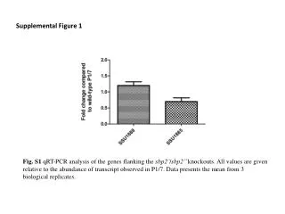
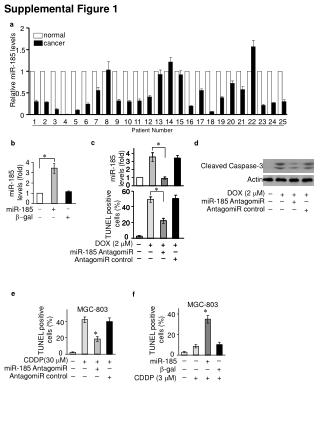
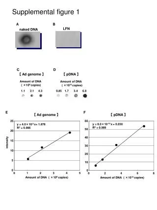
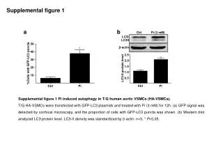
2.5 * 2.0 1.5 LC3-II protein level 1.0 0.5 Ctrl Pi Supplemental figure 1 a b Ctrl Pi (3 mM) LC3I LC3II 50 -actin * 40 30 %Cells with GFP-LC3 puncta 20 10 Ctrl Pi Supplemental figure 1 Pi induced autophagy in T/G human aortic VSMCs (HA-VSMCs). T/G HA-VSMCs were transfected with GFP-LC3 plasmids and treated with Pi (3 mM) for 12h. (a) GFP signal was detected by confocal microscopy, and the proportion of cells with GFP-LC3 puncta was shown. (b) Western blot analyzed LC3 protein level. LC3-II density was standardized by -actin. n=3, * P<0.05.
Supplemental figure 2 Anti-LC3 Von kossa staining Supplemental figure 2 LC3 puncta cells were found in both calcified and non-calcified aortic wall in chronic renal failure rats. Red arrows indicated autophagic cells, and red circle indicated calcified area.
Supplemental figure 3 1.5 1.0 Relative Atg5 mRNA level Supplemental figure 3Knockdown of Atg5 by siRNA. RatVSMCs were transfected with scramble or Atg5 small interfering RNA (siRNA) for 48 h. Quantitative real-time PCR analysis of Atg5 mRNA level. n=3, * P<0.05. 0.5 * Control Scramble Atg5 siRNA
Supplemental figure 4 Control Mn+Pi Mn+3-MA+Pi Pi 3-MA+Pi Supplemental figure 4 TUNEL staining (red) to identify apoptotic cells. Nuclei were counterstained with Hochest (blue). BASMCs were treated with 3-MA (5 mM), MnTMPyP (Mn, 25 M), and/or Pi (3 mM) as indicated for 7 days.
Supplemental figure 5 a b 8 50 # * * * * 40 6 * 30 MV ALP/Cell ALP ALP activity (U/ g prot) 4 20 2 10 Control Pi 3-MA 3-MA+Pi Control Pi 3-MA 3-MA+Pi Supplemental figure 5 The effects of autophagy inhibition on release of alkaline phosphatase (ALP) into the matrix vesicle (MV) fraction. (a) ALP activity of BASMCs after treatment with Pi (3 mM), 3-MA (5 mM) or Pi+3-MA for 3 days. (b) Data are expressed as the ratio of the cumulative release of MV ALP/total cellular ALP to indicate the effects of the drug on the release of ALP into the MV fraction. * P<0.05 vs control, # P<0.05 vs Pi.
SM--actin Cbf 1 Msx2 1.0 1.5 1.5 0.8 1.0 1.0 0.6 0.4 0.5 0.5 0.2 Supplemental figure 6 a SM22 * * 1.5 * * 1.0 Relative mRNA level Relative mRNA level Relative mRNA level Relative mRNA level 0.5 Ctrl Pi Ctrl Pi Ctrl Pi Ctrl Pi Ctrl Pi Ctrl Pi Ctrl Pi Ctrl Pi 3-MA 3-MA 3-MA Actin 3-MA Supplemental figure 6 The effects of autophagy inhibition on VSMC phenotypic transition. (a)Quantification of relative mRNA level of VSMC markers (SM22 and SM--actin) and osteogenic-related markers (core-binding factor 1 [Cbf1/Runx2] and msh homeobox 2 [Msx2]) in BASMCs treated with Pi (3 mM), 3-MA (5 mM) or Pi+3-MA for 3 days. n=4, * P<0.05.
Supplemental figure 6 continued Actin b Control Pi 3-MA Pi+3-MA SM--actin Supplemental figure 6 The effects of autophagy inhibition on VSMC phenotypic trasition. (b) Immunofluorescence distribution of actin and smooth muscle -actin (SM--actin) in BASMCs treated with Pi (3 mM), 3-MA (5 mM) or Pi+3-MA for 3 days. Red arrows showed the polarized distribution of actin in the perinuclear region in 3-MA treated VSMCs. White arrows showed the microvillar processes at the cellular edges that appeared to be devoid or greatly depleted of actin staining.