Download

1 / 29
490 likes | 2.36k Vues
Protein Turnover and Amino Acid Catabolism. Protein Degradation. Dietary Protein Digestion Cellular Protein Turnover. Dietary Protein Turnover. Proteins digested to amino acids and small peptides in the stomach Acid environment denatures proteins making them more accessible to proteases.

E N D
Protein Degradation • Dietary Protein Digestion • Cellular Protein Turnover
Dietary Protein Turnover • Proteins digested to amino acids and small peptides in the stomach • Acid environment denatures proteins making them more accessible to proteases. • Pepsin is a major stomach protease, has pH optimum of 2.0 • Protein degradation continues in the lumen of the intestine by pancreatic proteases • Amino acids are then released to the blood stream for absorption by other tissues.
Cellular Protein Turnover • Damaged proteins need to be degraded • Proteins involved in signaling are rapidly degraded to maintain tight regulation • Enzymes are often degraded as part of a pathway regulatory mechanism (HMG-CoA Reductase)
Protein Turnover Rates Vary • Proteins are constantly being degraded and resynthesized • Ornithine decraboxylase has short half life 11 minutes (polyamine synthesis-impt in cell growth and diff) • Hemoglobin and crystallin are very long lived protein • N-terminal amino acid residue determines protein stability
Lysosomal Hydrolysis • Proteins to be destroyed are encapsulated in vesicles • Proteins are deposited in lysosomes by the fusion of vesicles with the lysomomal membrane • Lysomomal proteases degrade protein.
Ubiquitin Related Protein Degradation • Ubiquitin is a small protein(8.5 kD = 76 amino acids) • Highly conserved among all Eukaryotes. • When covalently attached to a protein, ubiquitin marks that protein for destruction
Tagging of Proteins • The carboxyl-terminal glycine of ubiquitin covalently attaches to e-amino group of lysine residues on target protein • Requires ATP hydrolysis • Three enzymes involved: 1) E1, ubiqutiin activating protein, 2) E2, Ubiquitin conjugating enzyme, 3) E3, ubiquitin-protein ligase.
Protein Ubiquitination Multiple Ubiquitins can be polymerized to each other.
What determines whether a protein will become ubiquinated? • E3 enzyme are readers of N-terminal amino acid residues • N-terminal amino acids determine stability of protein • Also proteins rich in proline, glutamic acid, serine and threonine (PEST sequences) often have short ½ lives. • Other specific sequences (e.g. cyclin destruction box) target proteins for ubiquitination
Pathological Condition Related to Ubiquitination • Human papilloma virus encodes a protein that activates a specific form of the E3 enzyme that ubiquitinates several proteins involved in DNA repair. • Activation of this E3 enzyme is observed in 90% of cervical carcinomas.
Ubiquitinated Proteins are Degraded by the 26S Proteosome • The 26S proteosome is a large protease complex that specifically degrades ubiquinated proteins • 2 major components – 20S proteosome core, 19S cap. • Proteolysis occurs in 20S domain • Ubiquitin recognition occurs at 19S domain
26S Proteosome • ATP dependent process. • Protein is unfolded as it enters 20S domain. • Ubiquitin not degraded, but released and recycled.
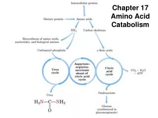
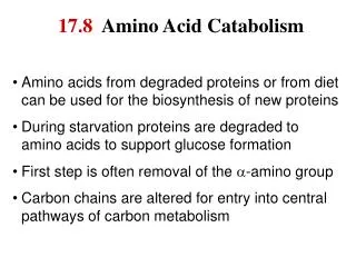
Fate of Amino Acids • Can be used for protein synthesis • If not needed for protein synthesis, must be degraded • In animals proteins and amino acids are not stored as a source of energy like can be carbohydrates and lipids. • Impt parts of amino acid degradation occur in the liver.
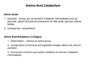
Amino Acid Catabolism • Deamination • Metabolism of Carbon Skeletons
Removal of nitrogen • Step 1: transamination with a-ketogluturate to form glutamate and new a-keto acid. • Step 2: glutamate is deaminated through oxidative process involving NAD+ • Step 3: form urea through urea cycle. deaminase transaminase
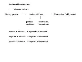
Fate of Ammonia • Ammonia (NH4+) is toxic. • Must not accumulate in cells. • In humans elevated levels are associated with lethargy and mental retardation • Mechanism of toxicity unknown.
Mechanisms to get rid of Ammonia • Fish excrete ammonia to aqueous environment through gills. • Birds and reptiles convert ammonia to uric acid and excrete it. • Mammals convert ammonia to urea in the liver and excrete it in urine. • Urea is soluble and uncharged, easy to excrete. Urea Uric Acid
Urea Cycle • 5 reaction cyclic pathway • Involves enzymes localized in the mitochondria and cytosol. • Two amino groups used derived from ammonia and aspartate. • C an O derived from bicarbonate
Step 1: Formation of Carbamoyl Phosphate • Reaction catalyzed by carbamoyl phosphate synthetase I • Most abundant enzyme in liver mitochondria (makes up 20% of matrix protein) • Allosterically activated by N-acetylglutamate (acetyl-CoA + glutamate N-acetylglutamate) • 2ATP + NH3 + Bicarbonate carbamoyl-P + 2ADP
Step 2: Ornithine Transcarbamyolase • Reaction occurs in mitochondrial matrix. • Product citrulline is exported out to cytosol
Step 3: Argininosuccinate Synthetase • Cytosolic enzyme • 2nd ammonia group incorporates from aspartate • ATP dependent reaction
Step 4: Argininosuccinase • Cytosolic enzyme
Step 5: Arginase • Cytosolic enzyme • Forms urea and ornithine. • Urea is excreted and ornithine is re-imported into mitochondria
Urea Cycle • Requires 3 ATPs + Ammonia + Aspartate + Bicarbonate • Get urea + fumurate + 2ADP + 2 Pi + AMP + PPi. • Fumurate skeleton feeds back into TCA
Glucose Alanine Cycle • Amino acid can be catabolized in muscle tissue where carbon skeletons are oxidized for energy. • Must remove toxic ammonia and transport to liver where it can be converted to urea. • Amino group from Glu is transferred to pyruvate to form alanine. • Alanine is exported to the liver via the blood stream where the it is deaminated to pyruvate • Pyruvate is converted to glucose which is returned to the muscle for fuel.