Download
1 / 28
300 likes | 663 Vues
D- Amino Acid Catabolism. A. Significance and Introduction B. Digestion C. Amino Transfer and Transport D. Nitrogen Excretion (Urea Cycle) E. Fate of the Carbon Skeletons. 6A. Significance (Table 18-2). Genetic Disorders Related to Amino Acid Metabolism

E N D
D- Amino Acid Catabolism A. Significance and Introduction B. Digestion C. Amino Transfer and Transport D. Nitrogen Excretion (Urea Cycle) E. Fate of the Carbon Skeletons
6A. Significance (Table 18-2) • Genetic Disorders Related to Amino Acid Metabolism • Most cases of genetic defects in amino acid metabolism lead to defective neural development* and mental retardation • (*Most a.a. are neurotransmitters, precursors, antagonists of) • Phenylketonuria (Phe hydroxylase mutation): phenylpyruvate and Phe accumulate in blood & are excreted in urine • Phe may compete with other amino acids for transport across blood-brain barrier à deficit of required metabolites • One of the first inheritable disease discovered in humans! • The disease can be controlled by a strict diet - avoiding slowed mental development
6A. Introduction • Energy gained from amino acids varies with each organism • Carnivores gain energy (up to 90%) from amino acid • oxidation • Plants rarely oxidize amino acids (CHO is energy source) • Amino acids can not be stored
Wild type Arabidopsis plant There are three reasons why amino acids need to be oxidatively degraded: 1. Protein turnover* 2. A high protein diet* 3. Starvation or diabetes (not enough CHO) *1 and 2 both involve an excess of amino acids Proteins are required for proper plant growth and development Mutant Arabidopsis plant has reduced levels of a regulator of protein degradation.
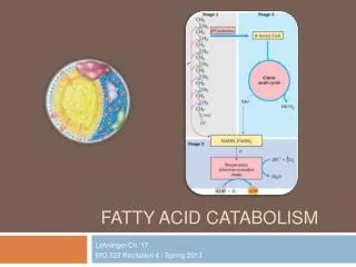
Overview of Amino Acid Catabolism in Mammals The amino groups and the carbon skeleton take separate but interconnected pathways
1. Protein 2. 3. 1. 4. 6B. Digestion 5. Cholecystokinin stimulates zymogen release 4. 6. 5. Low pH in small intestine stimulates secretin release and blood releases HCO3 7.
1. Protein enters stomach, stimulates gastrin secretion • 2. Gastrin (hormone) stimulates release of pepsinogen & HCl • 3. HCl is antiseptic and denatures proteins • Pepsinogen (activated by autoproteolytic cleavage at • lower pH) is the precursor of pepsin that cuts large • proteins into smaller peptide fragments and amino acids, • causing cholecystokinin release to the blood, stimulating • release of zymogens to the pancreas • 5. Low pH in the small intestine triggers secretin release in • blood, stimulating bicarbonate release (neutralize pH) • 6. Other proteases (neutral pH optimum) have been released • from the pancreas by the action of another hormone
Why synthesize the digestive enzymes as inactive precursors? Protects the exocrine cells from proteolytic attack 1. 2. (Found in intestine) 3. Figure 6-38 Note: this is a form of regulation … Biochemistry 221 - Metabolism
6B. Digestion • Trypsin can be inhibited in the pancreas by pancreatic • trypsin inhibitor • Digestion of proteins by trypsin and chymotrypsin is very fast since each has a different specificity • The carboxy- and aminopeptidases participate in degrading shorter peptides • Carboxy and amino terminal resides are removed one at a time • Once free, amino acids are transported through the intestinal mucosa through the blood and to the liver • Next we see what happens in the liver …
6C. Amino Group Transfer and Transport *Ammonia transport: Pyruvate (muscle) and glutamate can accept an amino group and are therefore suitable to carry ammonia through the blood. Process 1 Process 3 Process 2
“Glucose-Alanine Cycle” • Muscle protein is broken down to • amino acids that can be used for • fuel in muscle, where the end • product that contains the ammonia • is glutamate • Glu donates its amino group to • pyruvate through Ala • aminotransferase (next slide) to • produce Ala, and α-KG, which is • transported by the blood to the liver • In the liver, the reverse reaction • converts Ala and α-KG to pyruvate • that can be converted to glucose!
6C. Amino Group Transfer - Aminotransferase Purpose: to collect amino groups as L-Glu for excretion or biosynthetic pathways Process 1 & 2: Enzymatic removal of α-amino groups (transaminase /aminotransferases - named for amino donor i.e. Ala aminotranferase removes amino group from Ala) Keq’ ~ 1; ΔG'° ~ 0 kJ/mol - freely reversible reaction! Figure 18-4
6C. Amino Group Transfer - Aminotransferase • Process 1 & 2: Enzymatic removal of α-amino groups • Aminotransferases catalyze bimolecular Ping-Pong reaction where the first substrate reacts and the product leaves the active site before the second substrate binds • General Mechanism (see movies and next slides) • One amino acid binds, donates its amino group to PLP* and leaves as an α-keto acid • 2. Another α-keto acid binds and accepts the amino group from PLP, leaving as an amino acid • *Pyridoxal phosphate (PLP) acts as the amino group carrier
6C. Amino Group Transfer - Aminotransferase Role of PLP: Pyridoxal phosphate is the coenzyme form of pyridoxine - Vitamin B6 (deficiency leads to anemia) • Forms a Schiff • base with: • Lys of enzyme • Incoming a.a. • 3. Amino acid • donates amino • group with • release of • α-ketoacid (α-k.a.) • 4. Binding of new • α-k.a. and • release of a.a. bound coenzyme Aminotransferase Figure 18-5 Biochemistry 221 - Metabolism
6C. Amino Group Transfer - Aminotransferase Schiff Base Formation - Movie 18-1 Aminotransferase - Bimolecular Ping Pong Reaction
6C. Amino Group Transport – Gln Synthetase • Ammonia is toxic to animals and requires conversion before transport in the blood • Process 3: Transfer of free ammonia to L-Glu for transport in the blood as Gln: • L-Gln can also be used for biosynthetic reactions • Excess is transported to liver/kidney and deaminated by glutaminase (ammonia --> urea cycle) • Alanine can carry ammonia AND the carbon skeleton of pyruvate between liver and muscles Glutamine Synthetase ATP ADP + Pi
6C. Amino Group Transfer– Glu Dehydrogenase Once in the liver, amino groups are transferred to α-ketoglutarate to make L-Glu which is transported to the mitochondria for oxidative deamination (below): L-Glu DH uses NAD+ or NADP+ to accept reducing eq. Transdeamination: describes the combined action of the aminotransferase and Glu dehydrogenase. The α-ketoglutarate formed is fed into the citric acid cycle or glucose synthesis. Figure 18-7
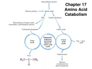
6C. Amino Group Transport and Transfer How is the Amino Group (or Ammonia) Used? 1. Most aquatic species (bony fish) secrete ammonia (ammonotelic) – toxic ammonia diluted by H2O 2. Plants recycle most of their ammonia. 3. Reptiles and birds excrete uric acid (uricotelic) 4. Synthesis of new amino acids or other products 5. Converted to urea (Figure 18-10) - most terrestrial animals secrete urea – this leads us to the Urea Cycle … (ureotelic)
6D. The Urea Cycle (Liver) • Chemical Origins of Urea • One NH2is derived from the deamination of Glu or Gln in • the mitochondria through carbamoyl phosphate • The central carbon is derived from bicarbonate, also through carbamoyl phosphate (synthesis, next slide) • The other NH2is donated by Asp
6D. The Urea Cycle • The activation of carbamoyl • phosphate requires 2 ATP • This reaction uses HCO3- • that is a byproduct of • mitochondrial respiration • *The carbamoyl phosphate • amino group originates from • ammonia that has been • transported to the liver • as 1. ammonia (portal vein), • 2. glutamine (other tissues), • 3. amino acids (glutamate) • or 4. alanine (muscle) *
6D. The Urea Cycle (Discovered by H. Krebs) Arg regulates Carbamoyl P Synthetase I Figure 18-9 Biochemistry 221 - Metabolism
6D. The Urea Cycle - almost exclusive to the liver • Some reactions happen in cytosol, others in mitochondria • Mitochondrial carbamoyl phosphate synthetase I is regulated • indirectly through Arg (N-acetylGlu and its synthetase) • Another ATP is used up in the conversion of citrulline to • argininosuccinate • Aspartate, required in the cytosolic conversion of citrulline • to argininosuccinate, is produced when oxaloacetate accepts an amino group from glutamate • None of the cytosolic components are part of the general • pool of cytosolic metabolites, since they are channeled between enzymes
6E. Fate of the Carbon Skeletons • Amino acids can account for 10-15% of the metabolic energy generated by mammals (omnivores) • All amino acids become CAC intermediates • CAC intermediates are: • 1. Diverted to gluconeogenesis (formation of glucose) • 2. Diverted to ketogenesis (formation of ketone bodies) • 3. Completely oxidized to CO2 and H2O
6E. Fate of the Carbon Skeletons ‡ * Ketone Bodies or Fatty Acids * *Glucogenic* ‡Ketogenic‡ * Glucose ‡ * ‡ * Figure 18-14, 18-29 Biochemistry 221 - Metabolism
6E. Fate of the Carbon Skeletons • The Cofactors • transfer one-carbon groups in different oxidation states • Tetrahydrofolate (folate vitamin): will transfer -CH3, - CH2OH or -(C=O)-H (at the 10N e.g. 10N-(C=O)-H) • 2. S-adenosylmethionine (SAM; Met + adenosine): is the preferred cofactor for methyl group transfers • 3. Biotin (previously discussed) – transfers CO2 • 4. Tetrahydrobiopterin: participates in redox reactions Figure 18-16
Glycinamide Ribonucleotide Transformylase uses THF! Interest slide
6E. Fate of the Carbon Skeletons In this figure, the red circles with red Xs indicate a genetic defect. Figure 18-22
6E. Fate of the Carbon Skeletons • Alternate pathways that • are driven when there is • a Phe build-up • All three products build • up in the tissues, blood and • urine • Phenylacetate produces an • odor in the urine that is • characteristic of phenyl- • ketonuria – if a nurse is • very observant, they will • notice this!