Download
1 / 50
500 likes | 607 Vues
This introduction to amino acid metabolism explores the synthesis and categorization of amino acids in human tissues, with a focus on the liver's crucial role in nitrogen metabolism. It explains the processes of transamination, deamination, and urea formation under dietary surplus, and differentiates between essential and non-essential amino acids. The metabolism pathways for glucogenic, ketogenic, and both types of amino acids are discussed, highlighting their roles in energy production and nitrogen elimination. The glucose-alanine cycle is also detailed, illuminating its significance for skeletal muscle.

E N D
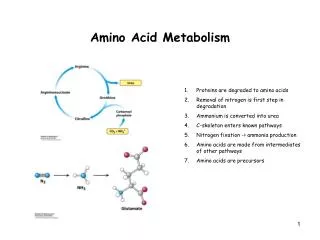
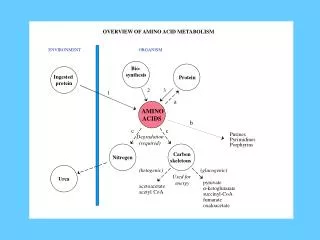
Introduction • All tissues have some capability for synthesis of the non-essential amino acids, amino acid remodeling, and conversion of non-amino acid carbon skeletons into amino acids and other derivatives that contain nitrogen. However, the liver is the major site of nitrogen metabolism in the body. In times of dietary surplus, the potentially toxic nitrogen of amino acids is eliminated via transaminations, deamination, and urea formation; the carbon skeletons are generally conserved as carbohydrate, via gluconeogenesis, or as fatty acid via fatty acid synthesis pathways. In this respect amino acids fall into three categories: glucogenic, ketogenic, or glucogenic and ketogenic. Glucogenic amino acids are those that give rise to a net production of pyruvate or TCA cycle intermediates, such as α-ketoglutarate or oxaloacetate, all of which are precursors to glucose via gluconeogenesis. All amino acids except lysine and leucine are at least partly glucogenic. Lysine and leucine are the only amino acids that are solely ketogenic, giving rise only to acetylCoA or acetoacetylCoA, neither of which can bring about net glucose production.
Introduction • A small group of amino acids comprised of isoleucine, phenylalanine, threonine, tryptophan, and tyrosine give rise to both glucose and fatty acid precursors and are thus characterized as being glucogenic and ketogenic. Finally, it should be recognized that amino acids have a third possible fate. During times of starvation the reduced carbon skeleton is used for energy production, with the result that it is oxidized to CO2 and H2O.
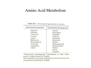
Essential vs. Nonessential Amino Acids • *The amino acids arginine, methionine and phenylalanine are considered essential for reasons not directly related to lack of synthesis. Arginine is synthesized by mammalian cells but at a rate that is insufficient to meet the growth needs of the body and the majority that is synthesized is cleaved to form urea. Methionine is required in large amounts to produce cysteine if the latter amino acid is not adequately supplied in the diet. Similarly, phenyalanine is needed in large amounts to form tyrosine if the latter is not adequately supplied in the diet.
Glutamate and Aspartate • Glutamate is synthesized from its' widely distributed α-keto acid precursor by a simple 1-step transamination reaction catalyzed by glutamate dehydrogenase. As discussed in the Nitrogen Metabolism page, the glutamate dehydrogenase reaction plays a central role in overall nitrogen homeostasis.
Reactions of glutamate dehydrogenase • Like glutamate, aspartate is synthesized by a simple 1-step transamination reaction catalyzed by aspartate aminotransferase, AST (formerly referred to as serum glutamate-oxalate transaminase, SGOT).
Aspartate can also be derived from asparagine (whose synthesis is outlined below) through the action of asparaginase. The importance of aspartate as a precursor of ornithine for the urea cycle is described in the Nitrogen Metabolism page.
Alanine and the Glucose-Alanine Cycle • Aside from its role in protein synthesis, alanine is second only to glutamine in prominence as a circulating amino acid. In this capacity it serves a unique role in the transfer of nitrogen from peripheral tissue to the liver. Alanine is transferred to the circulation by many tissues, but mainly by muscle, in which alanine is formed from pyruvate at a rate proportional to intracellular pyruvate levels. Liver accumulates plasma alanine, reverses the transamination that occurs in muscle, and proportionately increases urea production. The pyruvate is either oxidized or converted to glucose via gluconeogenesis. When alanine transfer from muscle to liver is coupled with glucose transport from liver back to muscle, the process is known as the glucose-alanine cycle. The key feature of the cycle is that in 1 molecule, alanine, peripheral tissue exports pyruvate and ammonia (which are potentially rate-limiting for metabolism) to the liver, where the carbon skeleton is recycled and most nitrogen eliminated.
There are 2 main pathways to production of muscle alanine: directly from protein degradation, and via the transamination of pyruvate by alanine transaminase, ALT (also referred to as serum glutamate-pyruvate transaminase, SGPT).
glucose-alanine cycle • The glucose-alanine cycle is used primarily as a mechanism for skeletal muscle to eliminate nitrogen while replenishing its energy supply. Glucose oxidation produces pyruvate which can undergo transamination to alanine. This reaction is catalyzed by alanine transaminase, ALT (ALT used to be called serum glutamate-pyruvate transaminase, SGPT). Additionally, during periods of fasting, skeletal muscle protein is degraded for the energy value of the amino acid carbons and alanine is a major amino acid in protein. The alanine then enters the blood stream and is transported to the liver. Within the liver alanine is converted back to pyruvate which is then a source of carbon atoms for gluconeogenesis. The newly formed glucose can then enter the blood for delivery back to the muscle. The amino group transported from the muscle to the liver in the form of alanine is converted to urea in the urea cycle and excreted.
Cysteine Biosynthesis • The sulfur for cysteine synthesis comes from the essential amino acid methionine. A condensation of ATP and methionine catalyzed by methionine adenosyltransferase yields S-adenosylmethionine (SAM or AdoMet).
Biosynthesis of S-adenosylmethionine, SAM • SAM serves as a precurosor for numerous methyl transfer reactions (e.g. the conversion of norepinephrine to epinenephrine, see Specialized Products of Amino Acids). The result of methyl transfer is the conversion of SAM to S-adenosylhomocysteine. S-adenosylhomocysteine is then cleaved by adenosylhomocyteinase to yield homocysteine and adenosine. Homocysteine can be converted back to methionine by methionine synthase, a reaction that occurs under methionine-sparing conditions and requires N5-methyl-tetrahydrofolate as methyl donor. This reaction was discussed in the context of vitamin B12-requiring enzymes in the Vitamins page. • Transmethylation reactions employing SAM are extremely important, but in this case the role of S-adenosylmethionine in transmethylation is secondary to the production of homocysteine (essentially a by-product of transmethylase activity). In the production of SAM all phosphates of an ATP are lost: one as Pi and two as PPi. It is adenosine which is transferred to methionine and not AMP. • In cysteine synthesis, homocysteine condenses with serine to produce cystathionine, which is subsequently cleaved by cystathionase to produce cysteine and α-ketobutyrate. The sum of the latter two reactions is known as trans-sulfuration.
Cysteine is used for protein synthesis and other body needs, while the α-ketobutyrate is first converted to propionyl-CoA and then via a 3-step process to the TCA cycle intermediate succinyl-CoA. While cysteine readily oxidizes in air to form the disulfide cystine, cells contain little if any free cystine because the ubiquitous reducing agent, glutathione, effectively reverses the formation of cystine by a non-enzymatic reduction reaction.
Utilization of Methionine in the Synthesis of Cysteine • The 2 key enzymes of this pathway, cystathionine synthase and cystathionase (cystathionine lyase), both use pyridoxal phosphate as a cofactor, and both are under regulatory control. Cystathionase is under negative allosteric control by cysteine, as well, cysteine inhibits the expression of the cystathionine synthase gene. • Genetic defects are known for both the synthase and the lyase. Missing or impaired cystathionine synthase leads to homocystinuria and is often associated with mental retardation, although the complete syndrome is multifaceted and many individuals with this disease are mentally normal. Some instances of genetic homocystinuria respond favorably to pyridoxine therapy, suggesting that in these cases the defect in cystathionine synthase is a decreased affinity for the cofactor. Missing or impaired cystathionase leads to excretion of cystathionine in the urine but does not have any other untoward effects. Rare cases are known in which cystathionase is defective and operates at a low level. This genetic disease leads to methioninuria with no other consequences. • Elevated levels of homocysteine in the blood have been shown to correlate with cardiovascular dysfunction. The role of homocysteine in cardiovascular disease is related to its ability to induce a state of inflammation. Homocysteine serves as a negatively charged surface that attracts the contact phase of the intrinsic pathway of blood coagulation. Activation of the intrinsic coagulation cascade leads to inappropriate thrombolytic events as well as resulting in increases in inflammatory cytokine release from leukocytes that are activated as a result of the pro-coagulant state. Therefore, it is important to ensure that proper function of the methionine synthase reaction is maintained. Although it would be assumed that increased intake of vitamin B12 should lead to increased conversion of homocysteine to methionine, and thus reduced levels of circulating homocysteine, controlled studies have shown that this does not occur.
Tyrosine Biosynthesis • Tyrosine is produced in cells by hydroxylating the essential amino acid phenylalanine. This relationship is much like that between cysteine and methionine. Half of the phenylalanine required goes into the production of tyrosine; if the diet is rich in tyrosine itself, the requirements for phenylalanine are reduced by about 50%. • Phenylalanine hydroxylase is a mixed-function oxygenase: one atom of oxygen is incorporated into water and the other into the hydroxyl of tyrosine. The reductant is the tetrahydrofolate-related cofactor tetrahydrobiopterin, which is maintained in the reduced state by the NADH-dependent enzyme dihydropteridine reductase (DHPR).
Biosynthesis of Tyrosine from Phenylalanine • Missing or deficient phenylalanine hydroxylase results in hyperphenylalaninemia. Hyperphenylalaninemia is defined as a plasma phenylalanine concentration greater than 2mg/dL (120μM). The most widely recognized hyperphenylalaninemia (and most severe) is the genetic disease known as phenlyketonuria (PKU). Patients suffering from PKU have plasma phenylalanine levels >1000μM, whereas the non-PKU hyperphenylalaninemias exhibit levels of plasma phenylalanine <1000μM. Untreated PKU leads to severe mental retardation. The mental retardation is caused by the accumulation of phenylalanine, which becomes a major donor of amino groups in aminotransferase activity and depletes neural tissue of α-ketoglutarate. This absence of α-ketoglutarate in the brain shuts down the TCA cycle and the associated production of aerobic energy, which is essential to normal brain development. • The product of phenylalanine transamination, phenylpyruvic acid, is reduced to phenylacetate and phenyllactate, and all 3 compounds appear in the urine. The presence of phenylacetate in the urine imparts a "mousy" odor. If the problem is diagnosed early, the addition of tyrosine and restriction of phenylalanine from the diet can minimize the extent of mental retardation. • Because of the requirement for tetrahydrobiopterin in the function of phenylalanine hydroxylase, deficiencies in DHPR can manifest with hyperphenylalaninemia. However, since tetrahydrobiopterin is a cofactor in several other enzyme catalyzed reactions (e.g. see the synthesis of the tyrosine- and tryptophan-derived neurotransmitters as well as nitric oxide in Specialized Products of Amino Acids), the effects of missing or defective DHPR cause even more severe neurological difficulties than those usually associated with PKU caused by deficient phenylalanine hydroxylase activity.
Ornithine and Proline Biosynthesis • Glutamate is the precursor of both proline and ornithine, with glutamate semialdehyde being a branch point intermediate leading to one or the other of these 2 products. While ornithine is not one of the 20 amino acids used in protein synthesis, it plays a significant role as the acceptor of carbamoyl phosphate in the urea cycle. Ornithine serves an additional important role as the precursor for the synthesis of the polyamines. The production of ornithine from glutamate is important when dietary arginine, the other principal source of ornithine, is limited.
Synthesis of Ornithine and Proline from Glutamic Semialdehyde • The fate of glutamate semialdehyde depends on prevailing cellular conditions. Ornithine production occurs from the semialdehyde via a simple glutamate-dependent transamination, producing ornithine. When arginine concentrations become elevated, the ornithine contributed from the urea cycle plus that from glutamate semialdehyde inhibit the aminotransferase reaction, with accumulation of the semialdehyde as a result. The semialdehyde cyclizes spontaneously to Δ1-pyrroline-5-carboxylate which is then reduced to proline by an NADPH-dependent reductase.
Serine Biosynthesis • The main pathway to de novo biosynthesis of serine starts with the glycolytic intermediate 3-phosphoglycerate. An NADH-linked dehydrogenase converts 3-phosphoglycerate into a keto acid, 3-phosphopyruvate, suitable for subsequent transamination. Aminotransferase activity with glutamate as a donor produces 3-phosphoserine, which is converted to serine by phosphoserine phosphatase. • As indicated below, serine can be derived from glycine (and visa versa) by a single step reaction that involves serine hydroxymethyltransferase and tetrahydrofolate (THF).
Glycine Biosynthesis • The main pathway to glycine is a 1-step reversible reaction catalyzed by serine hydroxymethyltransferase This enzyme is a member of the family of one-carbon transferases and is also known as glycine hydroxymethyltransferase. This reaction involves the transfer of the hydroxymethyl group from serine to the cofactor tetrahydrofolate (THF), producing glycine and N5,N10-methylene-THF.
Glycine produced from serine or from the diet can also be oxidized by glycine decarboxylase (also referred to as the glycine cleavage complex, GCC) to yield a second equivalent of N5,N10-methylene-tetrahydrofolate as well as ammonia and CO2.
Glycine is involved in many anabolic reactions other than protein synthesis including the synthesis of purine nucleotides, heme, glutathione, creatine and serine.
Aspartate/Asparagine and Glutamate/Glutamine Biosynthesis • Glutamate is synthesized by the reductive amination of α-ketoglutarate catalyzed by glutamate dehydrogenase; it is thus a nitrogen-fixing reaction. In addition, glutamate arises by aminotransferase reactions, with the amino nitrogen being donated by a number of different amino acids. Thus, glutamate is a general collector of amino nitrogen. Aspartate is formed in a transamination reaction catalyzed by aspartate transaminase, AST. This reaction uses the aspartate α-keto acid analog, oxaloacetate, and glutamate as the amino donor. Aspartate can also be formed by deamination of asparagine catalyzed by asparaginase.
Asparagine synthetase and glutamine synthetase, catalyze the production of asparagine and glutamine from their respective α-amino acids. Glutamine is produced from glutamate by the direct incorporation of ammonia; and this can be considered another nitrogen fixing reaction. Asparagine, however, is formed by an amidotransferase reaction.
Aminotransferase reactions are readily reversible. The direction of any individual transamination depends principally on the concentration ratio of reactants and products. By contrast, transamidation reactions, which are dependent on ATP, are considered irreversible. As a consequence, the degradation of asparagine and glutamine take place by a hydrolytic pathway rather than by a reversal of the pathway by which they were formed. As indicated above, asparagine can be degraded to aspartate.
Glutamine/Glutamate and Asparagine/Aspartate Catabolism • Glutaminase is an important kidney tubule enzyme involved in converting glutamine (from liver and from other tissue) to glutamate and NH4+, with the NH4+ being excreted in the urine. Glutaminase activity is present in many other tissues as well, although its activity is not nearly as prominent as in the kidney. The glutamate produced from glutamine is converted to α-ketoglutarate, making glutamine a glucogenic amino acid. Asparaginase (see above) is also widely distributed within the body, where it converts asparagine into ammonia and aspartate. Aspartate transaminates to oxaloacetate, which follows the gluconeogenic pathway to glucose. Glutamate and aspartate are important in collecting and eliminating amino nitrogen via glutamine synthetase and the urea cycle, respectively. The catabolic path of the carbon skeletons involves simple 1-step aminotransferase reactions that directly produce net quantities of a TCA cycle intermediate. The glutamate dehydrogenase reaction operating in the direction of α-ketoglutarate production provides a second avenue leading from glutamate to gluconeogenesis.
Alanine Catabolism • Alanine is also important in intertissue nitrogen transport as part of the glucose-alanine cycle (see above). Alanine's catabolic pathway involves a simple aminotransferase reaction that directly produces pyruvate. Generally pyruvate produced by this pathway will result in the formation of oxaloacetate, although when the energy charge of a cell is low the pyruvate will be oxidized to CO2 and H2O via the PDH complex and the TCA cycle. This makes alanine a glucogenic amino acid.
Arginine, Ornithine and Proline Catabolism • The catabolism of arginine begins within the context of the urea cycle. It is hydrolyzed to urea and ornithine by arginase. • Ornithine, in excess of urea cycle needs, is transaminated to form glutamate semialdehyde. Glutamate semialdehyde can serve as the precursor for proline biosynthesis as described above or it can be converted to glutamate. • Proline catabolism is a reversal of its synthesis process. • The glutamate semialdehyde generated from ornithine and proline catabolism is oxidized to glutamate by an ATP-independent glutamate semialdehyde dehydrogenase. The glutamate can then be converted to α-ketoglutarate in a transamination reaction. Thus arginine, ornithine and proline, are glucogenic.
Serine Catabolism • The conversion of serine to glycine and then glycine oxidation to CO2 and NH3, with the production of two equivalents of N5,N10-methyleneTHF, was described above. Serine can be catabolized back to the glycolytic intermediate, 3-phosphoglycerate, by a pathway that is essentially a reversal of serine biosynthesis. However, the enzymes are different. Although it has been demonstrated in mammals such as rodents and dogs that serine can be converted to pyruvate through a deamination reaction catalyzed by serine/threonine dehydratase, this enzyme is lacking in humans.
Threonine Catabolism • There are at least 3 pathways for threonine catabolism although only two are significant in humans. One involves a pathway initiated by threonine dehydrogenase yielding α-amino-β-ketobutyrate. The α-amino-β-ketobutyrate is either converted to acetyl-CoA and glycine, via the action of α-amino-β-ketobutyrate lyase, or it can spontaneously degrade to aminoacetone which is converted to pyruvate. • The second pathway utilizes serine hydroxymethyltransferase. As indicated above this enzyme belongs to a family of one-carbon transferases and is alternatively named glycine hydroxymethyltransferase and threonine aldolase. The products of this reaction are acetyl-CoA and glycine. The glycine can be converted to serine via the same enzyme and the serine is then catabolized as described above yielding pyruvate and NH4+. Thus, via this catabolic pathway threonine yields ketogenic and glucogenic byproducts. • The additional pathway involves serine/threonine dehydratase yielding α-ketobutyrate which is further catabolized to propionyl-CoA and finally the TCA cycle intermediate, succinyl-CoA. However, as indicated above, although the serine/threonine dehydratase enzyme is expressed in many mammalian species, it is not expressed in humans.
Glycine Catabolism • Glycine is classified as a glucogenic amino acid, since it can be converted to serine by serine hydroxymethyltransferase (see above), and serine can be converted back to the glycolytic intermediate, 3-phosphoglycerate or to pyruvate by serine/threonine dehydratase. Nevertheless, the main glycine catabolic pathway leads to the production of CO2, ammonia, and one equivalent of N5,N10-methyleneTHF by the mitochondrial glycine decarboxylase, also called the glycine cleavage complex, GCC (see above).
Cysteine Catabolism • There are several pathways for cysteine catabolism. The simplest, but least important pathway is catalyzed by a liver desulfurase and produces hydrogen sulfide, (H2S) and pyruvate. The major catabolic pathway in animals is via cysteine dioxygenase that oxidizes the cysteine sulfhydryl to sulfinate, producing the intermediate cysteinesulfinate. Cysteinesulfinate can serve as a biosynthetic intermediate undergoing decarboxylation and oxidation to produce taurine. Catabolism of cysteinesulfinate proceeds through transamination to β-sulfinylpyruvate which then undergoes desulfuration yielding bisulfite, (HSO3–) and the glucogenic product, pyruvate. The enzyme sulfite oxidase uses O2 and H2O to convert HSO3– to sulfate, (SO4–) and H2O2. The resultant sulfate is used as a precursor for the formation of 3'-phosphoadenosine-5'-phosphosulfate, (PAPS). PAPS is used for the transfer of sulfate to biological molecules such as the sugars of the glycosphingolipids.
Other than protein, the most important product of cysteine metabolism is the bile salt precursor taurine, which is used to form the bile acid conjugates taurocholate and taurochenodeoxycholate. The enzyme cystathionase can also transfer the sulfur from one cysteine to another generating thiocysteine and pyruvate. Transamination of cysteine yields β-mercaptopyruvate which then reacts with sulfite, (SO32–), to produce thiosulfate, (S2O32–) and pyruvate. Both thiocysteine and thiosulfate can be used by the enzyme rhodanese to incorporate sulfur into cyanide, (CN–), thereby detoxifying the cyanide to thiocyanate.
Methionine Catabolism • The principal fates of the essential amino acid methionine are incorporation into polypeptide chains, and use in the production of α-ketobutyrate and cysteine via SAM as described above. The transulfuration reactions that produce cysteine from homocysteine and serine also produce α-ketobutyrate, the latter being converted first to propionyl-CoA and then via a 3-step process to succinyl-CoA. • Regulation of the methionine metabolic pathway is based on the availability of methionine and cysteine. If both amino acids are present in adequate quantities, SAM accumulates and is a positive effector on cystathionine synthase, encouraging the production of cysteine and α-ketobutyrate (both of which are glucogenic). However, if methionine is scarce, SAM will form only in small quantities, thus limiting cystathionine synthase activity. Under these conditions accumulated homocysteine is remethylated to methionine, using N5-methylTHF and other compounds as methyl donors.
Valine, Leucine and Isoleucine Catabolism • This group of essential amino acids are identified as the branched-chain amino acids, BCAAs. Because this arrangement of carbon atoms cannot be made by humans, these amino acids are an essential element in the diet. The catabolism of all three compounds initiates in muscle and yields NADH and FADH2 which can be utilized for ATP generation. The catabolism of all three of these amino acids uses the same enzymes in the first two steps. The first step in each case is a transamination using a single BCAA aminotransferase, with α-ketoglutarate as amine acceptor. As a result, three different α-keto acids are produced and are oxidized using a common branched-chain α-keto acid dehydrogenase (BCKD), yielding the three different CoA derivatives. Subsequently the metabolic pathways diverge, producing many intermediates. • The principal product from valine is propionylCoA, the glucogenic precursor of succinyl-CoA. Isoleucine catabolism terminates with production of acetylCoA and propionylCoA; thus isoleucine is both glucogenic and ketogenic. Leucine gives rise to acetylCoA and acetoacetylCoA, and is thus classified as strictly ketogenic. • There are a number of genetic diseases associated with faulty catabolism of the BCAAs. The most common defect is in the branched-chain α-keto acid dehydrogenase, BCKD. Since there is only one dehydrogenase enzyme for all three amino acids, all three α-keto acids accumulate and are excreted in the urine. The disease is known as Maple syrup urine disease because of the characteristic odor of the urine in afflicted individuals. Mental retardation in these cases is extensive. Unfortunately, since these are essential amino acids, they cannot be heavily restricted in the diet; ultimately, the life of afflicted individuals is short and development is abnormal The main neurological problems are due to poor formation of myelin in the CNS.
Phenylalanine and Tyrosine Catabolism • Phenylalanine normally has only two fates: incorporation into polypeptide chains, and production of tyrosine via the tetrahydrobiopterin-requiring phenylalanine hydroxylase. Thus, phenylalanine catabolism always follows the pathway of tyrosine catabolism. The main pathway for tyrosine degradation involves conversion to fumarate and acetoacetate, allowing phenylalanine and tyrosine to be classified as both glucogenic and ketogenic. • Tyrosine is equally important for protein biosynthesis as well as an intermediate in the biosynthesis of several physiologically important metabolites e.g. dopamine, norepinephrine and epinephrine (see Specialized Products of Amino Acids). • As in phenylketonuria (deficiency of phenylalanine hydroxylase, PAH), deficiency of tyrosine aminotransferase (TAT) leads to hypertyrosinemia and the urinary excretion of tyrosine and the catabolic intermediates between phenylalanine and tyrosine. The adverse neurological symptoms are similar for PAH and TAT deficiencies. In addition, hypertyrosinemia leads to painful corneal eruptions and photophobia. • The first inborn error in metabolism ever recognized, alkaptonuria, was demonstrated to be the result of a defect in phenylalanine and tyrosine catabolism. Alkaptonuria is caused by defective homogentisic acid oxidase. Homogentisic acid accumulation is relatively innocuous, causing urine to darken on exposure to air, but no life-threatening effects accompany the disease. The only untoward consequence of alkaptonuria is ochronosis (bluish-black discoloration of the tissues) and arthritis.
Lysine Catabolism • Lysine catabolism is unusual in the way that the ε-amino group is transferred to α-ketoglutarate and into the general nitrogen pool. The reaction is a transamination in which the ε-amino group is transferred to the α-keto carbon of α-ketoglutarate forming the metabolite, saccharopine. Unlike the majority of transamination reactions, this one does not employ pyridoxal phosphate as a cofactor. Saccharopine is immediately hydrolyzed by the enzyme α-aminoadipic semialdehyde synthase in such a way that the amino nitrogen remains with the α-carbon of α-ketoglutarate, producing glutamate and α-aminoadipic semialdehyde. Because this transamination reaction is not reversible, lysine is an essential amino acid. The ultimate end-product of lysine catabolism is acetoacetyl-CoA. • Genetic deficiencies in the enzyme α-aminoadipic semialdehyde synthase have been observed in individuals who excrete large quantities of urinary lysine and some saccharopine. The lysinemia and associated lysinuria are benign. Other serious disorders associated with lysine metabolism are due to failure of the transport system for lysine and the other dibasic amino acids across the intestinal wall. Lysine is essential for protein synthesis; a deficiencies of its transport into the body can cause seriously diminished levels of protein synthesis. Probably more significant however, is the fact that arginine is transported on the same dibasic amino acid carrier, and resulting arginine deficiencies limit the quantity of ornithine available for the urea cycle. The result is severe hyperammonemia after a meal rich in protein. The addition of citrulline to the diet prevents the hyperammonemia.
Lysine is also important as a precursor for the synthesis of carnitine, required for the transport of fatty acids into the mitochondria for oxidation. Free lysine does not serve as the precursor for this reaction, rather the modified lysine found in certain proteins. Some proteins modify lysine to trimethyllysine using SAM as the methyl donor to transfer methyl groups to the ε-amino of the lysine side chain. Hydrolysis of proteins containing trimethyllysine provides the substrate for the subsequent conversion to carnitine.
Histidine Catabolism • Histidine catabolism begins with release of the α-amino group catalyzed by histidase, introducing a double bond into the molecule. As a result, the deaminated product, urocanate, is not the usual α-keto acid associated with loss of α-amino nitrogens. The end product of histidine catabolism is glutamate, making histidine one of the glucogenic amino acids. • Another key feature of histidine catabolism is that it serves as a source of ring nitrogen to combine with tetrahydrofolate (THF), producing the 1-carbon THF intermediate known as N5-formiminoTHF. The latter reaction is one of two routes to N5-formiminoTHF. Urocanate is converted to 4-imidazolone-5-propionate via the action of urocanate hydratase. The latter product is then converted to N-formiminoglutamte via the action of imidazolone propionase. Glutamate formiminotransferase then transfers the fomimino group to THF yielding glutamate and N5-formiminoTHF. • The principal genetic deficiency associated with histidine metabolism is absence or deficiency of the first enzyme of the pathway, histidase. The resultant histidinemia is relatively benign. The disease, which is of relatively high incidence (1 in 10,000), is most easily detected by the absence of urocanate from skin and sweat, where it is normally found in relative abundance.
Decarboxylation of histidine in the intestine by bacteria gives rise to histamine. Similarly, histamine arises in many tissues by the decarboxylation of histidine, which in excess causes constriction or dilation of various blood vessels. The general symptoms are those of asthma and various allergic reactions. Synthesis of Histamine
Tryptophan Catabolism • A number of important side reactions occur during the catabolism of tryptophan on the pathway to acetoacetate. The first enzyme of the catabolic pathway is an iron porphyrin oxygenase that opens the indole ring. The latter enzyme is highly inducible, its concentration rising almost 10-fold on a diet high in tryptophan. • Kynurenine is the first key branch point intermediate in the catabolic pathway leading to 3 fates: • Kynurenine can undergo deamination in a standard transamination reaction yielding kynurenic acid. Kynurenic acid and metabolites have been shown to act as antiexcitotoxics and anticonvulsives. High levels of kynurenic acid have been found in the urine of individuals suffering from schizophrenia. Kynurenic acid has been shown to act as a non-competetive antagonist at the glycine binding site of the NMDA receptor (NMDA = N-methyl-D-aspartate) which is an ionotropic (ligand-gated ion channel) receptor for glutamate. The NMDA receptor is a key component of the glutaminergic neurotransmission system believed to be involved in the pathophysiology of schizophrenia, thus explaining the potential role of kynurenic acid in schizophrenia.
Kynurenine can also undergo a series of catabolic reactions producing 3-hydroxyanthranilic acid plus alanine. Another equivalent of alanine is produced from kynurenine in a single step reaction leading to anthranilic acid. It is the production of these alanine residues that allows tryptophan to be classified among the glucogenic amino acids. Oxidation of 3-hydroxyanthranilate converts it into 2-amino-3-carboxymuconic 6-semialdehyde, which has two fates. The main flow of carbon elements from this intermediate leads to acetoacetate which is why tryptophan is also a ketogenic amino acid. An important side reaction in liver involves a non-enzymatic cyclization to quinolate then via a transamination and several rearrangements yields limited amounts of nicotinic acid, which leads to production of a small amount of NAD+ and NADP+. • Aside from its role as an amino acid in protein biosynthesis, tryptophan also serves as a precursor for the synthesis of serotonin and melatonin. These products are discussed in Specialized Products of Amino Acids.