Cell Structures and Functions: An In-depth Study
Explore the diverse world of cell structures, from prokaryotes to eukaryotes, membranes to organelles. Learn about size limitations, endomembrane system, wall composition, and more. Engage in discussions on cell types and junctions.

Cell Structures and Functions: An In-depth Study
E N D
Presentation Transcript
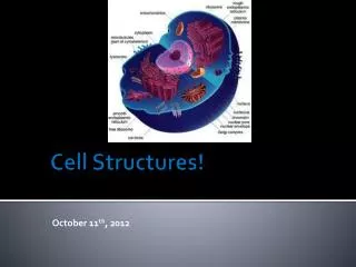
Cell Structures 2 Slides to be used for Class discussion
Cell size is limited by two factors The surface area increases at a slower rate than the volume for a sphere or cube shape. The larger the cell the longer it takes for materials to diffuse to the center of the cell
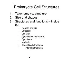
Prokaryotes - Basics • Plasma membrane • Ribosomes • Cell wall • 1 chromosome in nucleoid region • Small size (0.5-2μm)
Inside Prokaryotes • Limited cytoskeleton • Many have compartments/ organelles • Many have plasmids • Small circular DNA • Many have photosynthetic membranes (lamella)
Outside Prokaryotes • Some have flagella • Most have a cell wall • Made of peptidoglycans (eubacteria) or polysaccharides (archaebacteria) • Some have an additional outer capsule composed of glycolipids (protection, adhere)
Eukaryotes - Basics More highly evolved and much larger than prokaryotes (10-100μm) Chromosomes within a nucleus Network of cytoskeleton Numerous membrane-bound organelles to separate chemical reactions Evolved from involution of the membrane Prokaryotic cells
The signal hypothesis predicts that proteins bound for the endomembrane system have a “zip code” that directs the growing polypeptide to the ER. • This “zip code” is a 20-amino-acid-long ER signal sequence. • The ER signal sequence binds to a signal recognition particle (SRP) that then binds to a receptor in the ER membrane.
Each packaged protein that leaves the Golgi has a molecular tag that places it in a particular type of transport vesicle, depending on its destination.
Vacuoles • Large storage structures • Store water, ions, nutrients • Help maintain cell rigidity (turgor pressure in plants) • Some contain digestive enzymes
Endosymbiosis Hypothesis • Mitochondria, chloroplasts, and the flagella evolved from prokaryotic cells engulfing other prokaryotic cells
The Cell Wall • Support • Not in animals • Eukaryote cell wall: made of cellullose (plants and algae) or chitin (fungi) • Some plants have a secondary cell wall containing lignin (in wood)
Extracellular Matrix • Outside animal cells • Consists of • Fibrous structural proteins • Collagen • Adhesion proteins • Link ECM to cytoskeleton • Polysacchardies • Structural support • Allow cells to stick together or to a substrate
Cytoskeleton • Shape and structure • Movement (internal and external) • Made of protein fibers • Microfilaments • Intermediate filaments • Microtubules
Microfilaments Smallest, made of actin Interact with myosin to change cell shape or bear tension Found in muscle cells, phagocytes, during cytokinesis
IntermediateFilaments Fibrous proteins Structure, no movement Flexible; help shape the cell surface and anchor nucleus
Microtubules • Large hollow tubes made of tubulin • Stability and movement • Polar (+ & - ends) • Originate from microtubule organizing center, grow out from their + ends • Called the centrosome (2 centrioles) in animals, 9 triplets of microtubules
Microtubules and Vesicle Transport • Requires ATP • Kinesin: a motor protein that converts chemical energy in ATP into mechanical work • Dynein: retrograde motor protein
Microtubules in Cilia and Flagella • Flagella • Long “tail” for motility • Bacteria • Made of flagellin • Propeller • Eukaryotes • Made of microtubules • Wave back and forth • Cilia • Short, filament-like projections
Cilia and Flagella Axoneme • Complex “9 + 2” arrangement of microtubules connected by links and spokes • Dynein protein found in links
Dynein • Changes shape when ATP is hydrolyzed to “walk” up the microtubule • The dynein movement alternates per side of axoneme • Cause cilia and flagella to bend
Cell Junctions • Anchor cells to one another • Provide a passageway for cellular exchange • Include • Anchoring junctions • Tight junctions • Communicating junctions • Gap junctions • Plasmodesmata
Epithelium in Animal Cells • Composed of sheets of cells that cover organs and line body cavities • Many types of structures connect neighboring epithelial cells, including tight junctions and desmosomes.
Tight Junctions • Animal cells • Specialized proteins in the plasma membranes • Stitch cells together to form a watertight seal • Found between cells in tissues that form a barrier (lining the digestive tract) • Force materials through cells (rather than intracellular spaces)
Desmosomes Proteins that link the cytoskeletons of adjacent cells Epithelial and muscle tissue These proteins bind to each other and to the proteins that anchor cytoskeletal intermediate filaments
Communicating Junctions • Direct connections between cells in the same tissue allow cells to communicate and work together in a coordinated fashion
Plasmodesmata • Connect plant cells • Gaps in the cell wall where the plasma membranes, cytoplasm, and smooth ER of two cells connect.
Gap Junctions • Connect animal tissues • Form channels that allow the flow of small molecules between cells.