Download
1 / 12
210 likes | 989 Vues
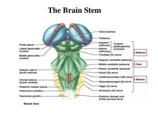
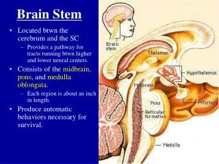
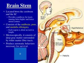
How to draw different sections of the brain stem. Sanjaya Adikari Department of Anatomy. Posterior surface. Anterior surface. Main nerve tracts in the spinal cord. B. A. C. D. G. E. F. Posterior white columns Lateral corticospinal tract Anterior & posterior spinocerebellar tracts

E N D
How to draw different sections of the brain stem Sanjaya Adikari Department of Anatomy
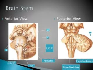
Posterior surface Anterior surface
Main nerve tracts in the spinal cord B A C D G E F
Posterior white columns Lateral corticospinal tract Anterior & posterior spinocerebellar tracts Anterior & posterior spinothalamic tracts Olivospinal, vestibulospinal, tectospinal tracts Anterior corticospinal tract Rubrospinal tract
Closed medulla at the level of decussation of pyramids Fasciculus gracilis Spinal tract of trigeminal nerve Nucleus gracilis Fasciculus cuneatus Nucleus cuneatus Spinal nucleus of trigeminal nerve Spinal root of the accessory nerve Posterior spinocerebellar tract Central canal Spinothalamic tract Medial longitudinal fasciculus Anterior spinocerebellar tract Decussation of pyramids Pyramid
Fasciculus gracilis Spinal tract of trigeminal nerve Nucleus gracilis Fasciculus cuneatus Nucleus cuneatus Spinal nucleus of trigeminal nerve Central canal Medial longitudinal fasciculus Posterior spinocerebellar tract Spinal root of the accessory nerve Spinothalamic tract Decussation of medial lemnisci Anterior spinocerebellar tract Pyramid Hypoglossal nerve Closed medulla at the level of decussation of medial lemnisci
Medulla Oblongata at the level of middle of the olivary nuclei Inferior medullary velum Cavity of 4th ventricle Medial longitudinal fasciculus Spinal tract & nucleus of trigeminal nerve Vestibular & cochlear nuclei Inferior cerebellar peduncle Vagus nerve Anterior spinocerebellar tract Reticular formation Spinothalamic tract Olivary nucleus Tectospinal tract Olive Hypoglossal nerve Medial lemniscus Pyramid
Pons at the level of facial colliculus Superior medullary velum Cavity of 4th ventricle Medial longitudinal fasciculus Superior cerebellar peduncle Facial colliculus Reticular formation Vestibular nuclei Inferior cerebellar peduncle Spinal lemniscus Spinal tract & nucleus of trigeminal nerve Medial lemniscus Transverse pontine fibres Facial nerve Abducent nerve Corticospinal & corticonuclear fibres Pontine nuclei
Midbrain at the level of inferior colliculus Cerebral aqueduct Trochlear nerve Periaqueductal Gray Matter Inferior colliculus Mesencephalic nucleus of trigeminal nerve Trigeminal lemniscus Spinal lemniscus Medial longitudinal fasciculus Medial lemniscus Substantia nigra Reticular formation Cerebral peduncle Decussation of superior cerebellar peduncles Interpeduncular fossa
Midbrain at the level of superior colliculus Cerebral aqueduct Periaqueductal Gray Matter Superior colliculus Mesencephalic nucleus of trigeminal nerve Trigeminal lemniscus Spinal lemniscus Medial longitudinal fasciculus Medial lemniscus Substantia nigra Reticular formation Cerebral peduncle Red nucleus Oculomotor nerve
PPRF = Paramedian pontine reticular formation Medial longitudinal fasciculus