Download
1 / 6
60 likes | 182 Vues
This study explores whether voltage-gated ionic flux can enhance acidification in macrophage endosomes during infection. Utilizing flow cytometry on isolated endosomes, we aim to complement microscopy and electrophysiology data. The experimental approach includes differentiating and priming a THP-1 monocytic cell line, isolating endosomes via sucrose gradient, dialyzing samples, and loading them with functional fluorescent dyes. We will analyze activated channels on a MoFlo flow cytometer to collect and study the enriched endosomal fraction while addressing technical challenges like particle size and pressure.

E N D
Functional Analysis of Ionic Flux in Isolated Endosomes Michael Carrithers Department of Neurology
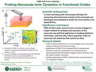
Can voltage-gated ionic flux enhance acidification of macrophage endosomes during infection?
Goal: Use flow cytometry on isolated endosomes to complement microscopy and electrophysiology.
Experimental Approach • Differentiate and prime a monocytic cell line (THP-1). • Isolate endosomes on a sucrose gradient. • Dialyze sample. • Load with functional fluorescent dyes. • Activate channel. • Analyze on MoFlo. • Collect enriched endosomal fraction.
Fluorescent Dyes • Lysosensor Green (pH) • Sodium Green • CoroNa Red • Disbac2(3) (slow voltage-sensitive dye)
Technical Challenges • Particle size (200-800 nm) • Pressure • Sheath buffer • Large amount of starting material