
Chapter 4
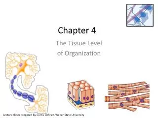
Chapter 4. The Tissue Level of Organization. Lecture slides prepared by Curtis DeFriez , Weber State University. Tissues. Tissues are a group of cells with a common embryonic origin that function together to carry out specialized activities. They include various types,

Chapter 4
E N D
Presentation Transcript
Chapter 4 The Tissue Level of Organization Lecture slides prepared by Curtis DeFriez, Weber State University
Tissues • Tissues are a group of cells with a common embryonic origin that function together to carry out specialized activities. • They include various types, ranging from hard (bone) to semisolid (fat) to liquid (blood).
Tissues • Histology is the study of the microscopic anatomy of cells and tissues – it is a branch of pathology. • Of the 10 trillion cells in our body, no single cell type can said to be “typical”. A trained histologist can recognize over 200 distinct human cell types under the microscope and is able to distinguish a cell from pancreatic tissue as opposed to a cell from the skin. • Each cell type has features particular to its function.
Intracellular Junctions • Tissues are formed by grouping cells together using a variety of Intercellular Junctions . • Intracellular Junctions connect adjacent cells mechanically at the cell membranes or through cytoskeletal elements within and between cells.
Intracellular Junctions • Tight Junctions are found where a leakproof seal is needed between cells. • They keep materials from leaking out of organs like the stomach and bladder.
Intracellular Junctions • Adherens Junctions make an adhesion belt (like the belt on your pants) that keeps tissues from separating as they stretch and contract. • Cadherin is a glycoprotein that forms the belt-like “plaque”.
Intracellular Junctions • Desmosomes act as “spot welds”. They also use cadherin glycoprotein (plus intermediate filaments) to hook into the cytoplasm.
Intracellular Junctions • Hemidesmosomes are half-welds that join cells to the basement membrane.
Intracellular Junctions • Gap Junctions are pores (connexons) that allow small substances like ions to pass between cells. If one of the cells gets sick or dies, these seal like a hatch to prevent damage to other cells.
Intracellular JunctionsInteractions Animation • Intracellular Junctions You must be connected to the internet to run this animation
The 4 Basic Tissues • Of all the cells in the body, they combine to make only 4 basic tissue types: • Epithelial tissues • Connective tissues • Muscular tissues • Nervous tissues
The 4 Basic Tissues • Epithelial tissues cover body surfaces and form glands and line hollow organs, body cavities, and ducts.
The 4 Basic Tissues • Connective tissues (C.T.) protect, support, and bind organs. • Fat is a type of C.T. that stores energy. • Red blood cells, white blood cells, and platelets are all C.T.
The 4 Basic Tissues • Muscular tissues generate the physical force needed to make body structures move. They also generate heat used by the body. • Nervous tissues detect changes in the body and respond by generating nerve impulses.
The 4 Basic Tissues • Tissues of the body develop from three primary germ layers: Endoderm, Mesoderm, and Ectoderm • Epithelial tissues from all three germ layers • C.T. and muscle are derived from mesoderm. • Nervous tissue develops from ectoderm.
Epithelium • Epithelium is used to line surfaces and form protective barriers. Epithelium is also good at secreting things like mucous, hormones, and other substances . • All epithelia have a free apical surface and an attached basal surface.
Epithelium • The basal layer of the epithelium secretes a basal lamina; the underlying C.T. secretes a reticular lamina. • Together the basal lamina and the reticular lamina form a non- cellular basement membrane on which the epithelium sits.
Epithelium • Epithelia are named according to the shape of their cells, and the thickness or arrangement of their layers (of cells).
Epithelium • Naming epithelia according to shape
Epithelium • Naming epithelia according to arrangement
Epithelium • Naming epithelia • Three different cell shapes x three different cell arrangements = nine possibilities. Two of these are not used. Add transitional (cells that change shape), and we’re back up to eight possible combinations. • If different shapes are present in layers of cells, the epithelium is always named by the shape of cells in the apical (outermost) layer.
Epithelium • Simple Squamous Epithelium is composed of a single layer of flat cells found: • In the air sacs of lungs • In the lining of blood vessels, the heart, and lymphatic vessels • In all capillaries, including those of the kidney • As the major part of a serous membrane
Epithelium • Simple Cuboidal Epithelium is composed of a single layer of cube shaped cells. • It is often found lining the tubules of the kidneys and many other glands.
Epithelium • Simple Columnar Epithelium forms a single layer of column-like cells, ± cilia, ± microvilli, ± mucous (goblet cells). • Goblet cells are simple columnar cells that have differentiated to acquire the ability to secrete mucous.
Epithelium • Pseudostratified Columnar Epithelium appears to have layers, due to nuclei which are at various depths. In reality, all cells are attached to the basement membrane in a single layer, but some do not extend to the apical surface. • Ciliated tissue has goblet cells that secrete mucous.
Epithelium • Stratified Squamous Epithelium has an apical surface that is made up of squamous (flat) cells. • The other layers have different shapes, but the name is based on the apical layer. • The many layers are ideal for protection against strong friction forces.
Epithelium • Stratified Cuboidal Epithelium has an apical surface made up of two or more layers of cube-shaped cells. • Locations include the sweat glands and part of the ♂ urethra • Stratified Columnar Epithelium is very rare, and for our purposes, hardly worth mentioning.
Epithelium • The cells of Transitional Epithelium change shape depending on the state of stretch in the tissue. • The apical “dome cells” of the top layer (seen here in relaxation) are an identifiable feature and signify an empty bladder . • In a full bladder, the cells are flattened.
Epithelium • Although epithelia are found throughout the body, certain ones are associated with specific body locations. • Stratified squamous epithelium is a prominent feature of the outer layers of the skin.
Epithelium • Simple squamous makes up epithelial membranes and lines the blood vessels. • Columnar is common in the digestive tract. • Pseudostratified ciliated columnar is characteristic of the upper respiratory tract. • Transitional is found in the bladder. • Cuboidal lines ducts and sweat glands.
Covering and Lining Epithelium • Endothelium is a specialized simple squamous epithelium that lines the entire circulatory system from the heart to the smallest capillary – it is extremely important in reducing turbulence of flow of blood. • Mesothelium is found in serous membranes such as the pericardium, pleura, and peritoneum. • Unlike other epithelial tissue, both are derived from embryonic mesoderm (the middle layer of the 3 primary germ layers of the embryo).
Connective Tissue • Connective Tissues are the most abundant and widely distributed tissues in the body – they are also the most heterogeneous of the tissue groups. • They perform numerous functions: • Bind tissues together • Support and strengthen tissue • Protect and insulate internal organs • Compartmentalize and transport • Energy reserves and immune responses
Connective Tissues • Collagen is the main protein of C.T. and the most abundant protein in the body, making up about 25% of total protein content. • Connective tissue is usually highly vascular and supplied with many nerves. • The exception is cartilage and tendon - both have little or no blood supply and no nerves.
Connective Tissues • Although they are a varied group, all C.T. share a common “theme”: • Sparse cells • Surrounded by an extracellular matrix • The extracellular matrix is a non-cellular material located between and around the cells. • It consists of protein fibers and ground substance (the ground substance may be fluid, semifluid, gelatinous, or calcified.)
Cells Of Connective Tissues • Common C.T. cells • Fibroblasts are the most numerous cell of connective tissues. These cells secrete protein fibers (collagen, elastin, & reticular fibers) and a “ground substance” which varies from one C.T. to another.
Cells of Connective Tissues • Of the other common C.T. cells: • Chondrocytes make the various cartilaginous C.T. • Adipocytes store triglycerides. • Osteocytes make bone. • White blood cells are part of the blood.
Connective Tissues • There are 5 types of white blood cells (WBCs): • Macrophages are the “big eaters” that swallow and destroy invaders or debris. They can be fixed or wandering. • Neutrophils are also macrophages (“small eaters”) that are numerous in the blood. • Mast cells and Eosinophils play an important role in inflammation. • Lymphocytes secrete antibody proteins and attack invaders.
Connective Tissues • C.T. cells secrete 3 common fibers: • Collagen fibers • Elastin fibers • Reticular fibers
Connective Tissues • This graphic represents a collage of different C.T. elements (cells and fibers) and not a specific C.T.


