Download
1 / 18
240 likes | 322 Vues
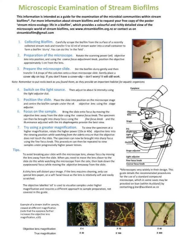
General Urine Examination Microscopic examination of urine. Advanced laboratory techniques Third stage Lecture (4) ( Theortical ) Asisstant lecturer Rajaa Saihood Abbas. Microscopic examination of urine.

E N D
General Urine Examination Microscopic examination of urine Advanced laboratory techniques Third stage Lecture (4) ( Theortical) Asisstant lecturer RajaaSaihood Abbas
Microscopic examination of urine • Urine is examined microscopically as a wet preparation to detect :- • 1-Significant pyuria ,i.e WBC in excess of 10 cells/microleter of urine • 2-Red cells • 3- Casts and • 4- Yeast cells • 5-T .vaginalismotile trophozoite • 6-S.haematobium egg and microfilaria • 7- Bacteria • 8- Epithelial cells • 9- Spermatozoa • 10- Crystals.
Microscopic examination of urine • For routine work as well as special request work with urine sample is divided into 2 tubes ; one portion is used without centrifugation(un centrifuged urine and the other portion is centrifuged. • Un centrifuged urine is used for:- • 1- Chemical & biochemical analysis • 2- Microscopic estimation of bacteria (bacteriuria) * • 3- Microscopic estimation of pyuria(WBCs) is the condition of urine containing white blood cells or pus. * • 4- Microscopic estimation of haematuria(RBC) * • 5- Estimation of haemoglobinuria (Hb) and occult blood.* • 6- Pregnancy tests. • 7- Detection of crystals • 8- Bacterial culture &gram staining,.
Microscopic examination of urine • While the centrifuged urine is used for:- • 1- Microscopic examination of bacteria. * • 2- Microscopic detection of casts. • 3- - Microscopic detection of parasite e.g. Schistosoma. Haematobium and Trichomonas .vaginalistrophozoite . • 4- Microscopic estimation of pyuria(WBCs) * • 5- Microscopic detection of Spermatozoa. • 6- Microscopic estimation of epithelial cells. • 7- Estimation of significant haematuria. * • 8- Gram staining for estimation of bacteria (bacteriuria
White blood cells (pus cells) • WBCs usually not found in normal urine (or very few)but their number increased in UTIs caused by many pathogens and called pyuria . • When we examined centrifuged urine(the sediments) WBCs are reported as:- • Few : up to 10 WBCs/HPF(high power field)using 40X objective lens • Moderate : 11-40 WBCs/HPF • Many : more than 40 WBCs/HPF • Pyuria is usually regarded as significant when moderate or many pus cell are present (more than 10 WBCs/HPF)in most UTIs caused by E.coli ,proteus ,klebsiella ,Staph ,Pseudomonuas , Leptospira , Clamydia and many others .However in some other conditions like enteric fever, UTI in diabetic people.
Sterile Pyuria Sterile Pyuria: is urine which contains white blood cells while appearing sterile by standard culturing techniques. It is often caused by sexually transmitted infections, such as gonorrhea, or viruses which will not grow in bacterial cultures. Sterile pyuria is listed as a side effect from some medications such as paracetamol (acetaminophen). Its occurrence is also associated with certain disease processes, such as Kawasaki disease and genitourinary tuberculosis.
Red blood cells Normally no blood is found in urine except in certain physiological conditions Like during menstruation period. Hematuria is the presence of red blood cells (erythrocytes) in the urine. It may be idiopathic or benign, or it can be a sign that there is a kidney stone or a tumor in the urinary tract (kidneys, ureters, urinary bladder, prostate, and urethra),urinary Schistosomiasis, acute glomerulonephritis, sickle cell disease Leptospirosis and hemorrhagic conditions.
Haemoglobinuria • Haemoglobinuria is a condition in which the oxygen transport protein hemoglobin is found in abnormally high concentrations in the urine. The condition is often associated with any hemolytic anemia with primarily intravascular hemolysis, in which red blood cells (RBCs) are destroyed, thereby releasing free hemoglobin into the plasma. Excess hemoglobin is filtered by the kidneys, which excrete it into the urine.
Bilirubin Bilirubin is a break down products of Hb . • Most of the bilirubin produced in humans is conjugated by the liver and excreted into the bile, but a very small amount of conjugated bilirubin is reabsorbed and reaches the general circulation to be excreted in the urine. • Bilirubin in the urine ( +ve test)indicate hepatic disease or hepato-biliary obstruction.
Urobilinogen • Urobilinogenis a substance formed in the gastro intestinal tract by the bacterial reduction of conjugated bilirubin. Increased urinary urobilinogen occurs in prehepatic jaundice(hemolytic anemia), hepatitis , hepatic necrosis and others .
Casts • These are masses of solidified protein or cells with cylindrical shape because they are formed in kidney tubules .Mostly they can be seen in centrifuged urine with 10 x lens .Different types of cast are seen in urine . • 1-Hyaline casts :-colorless empty masses indicates damage of glomeruli • 2- Waxy casts :- they are thicker and denser with twisted shapes indicate es tubules damage and renal failure. • 3-Cellular cast :- • a- RBC cast = orange color indicate haemorrhage in the renal tubules and glomerular bleeding . • b-WBC cast indicate pelvic or tubules infection (UTI) • 4- Granular cast which contain irregular granules originated from degenerated cells and protein and associated with renal damage
Yeast cells • Yeast cells can be seen in urine of women with vaginal candidiasis . • Parasites • 1- Trichomonas .vaginalistrophozoite is one of the causative agents of vaginitis in women(occasionally seen in the urine of men).The protozoa has only trophozoite from which can be detected in urine by its shape (bear-shape)or characteristic motility (corkscrew movement). • 2- Schistosoma .haematobium eggs • Eggs of S. haematobium can be excreted in urine after about 4-6 weeks of infection ,it can be detected in centrifuged urine or last drops of urine sample by its characteristic shape and size.
Microfilaria • 3- Microfilaria • Other parasites that can be detected in urine sample include:- • a- Microfilaria of Wuchereria .bancrofti can be occasionally seen in urine in case of lymphatic Microfilaria when lymphatic rupture occurs in the lymphatic vessels of urogenital system . • b- Microfilaria of Onchocerca. Volvulus can be detected in urine from patients with onchocerciasis . • 4- Occasionally the eggs of Entrobius .vermicularis are found in urine, especially from young girls when the eggs are washed off the external genitalia .
Epithelial cells • The tissue that lines the surfaces of cavities and structures in your body is called epithelial tissue. In healthy individuals, epithelial cells from the bladder and external urethra are normally present in the urine in small amounts. However, the amount of epithelial cells in the urine increases when someone has a urinary tract infection or some other cause of inflammation. Your doctor will evaluate the source of the problem by evaluating the type of epithelial cells that are present. For example, the presence of renal tubular epithelial cells (from your kidneys) may indicate kidney involvement. The presence of squamous epithelial cells may indicate vaginal contamination of the urine specimen.
Spermatozoa • Occasionally found in the urine of men or women after sex intercourse • Bacteria: • Bacteria in your urine can suggest infection, especially if you have other suggestive symptoms. If your doctor suspects that you may have a urinary tract infection, she/he will most likely order a culture or count of the bacteria, Gram stain is recommended to identify primary the shape of bacteria ,usually seen as rods ,coccid or streptococci . However, bacteria on the skin can also contaminate the urine sample and skew the results, so it is very important that you understand proper aseptic technique when giving a urine sample.
Crystals Crystals can be present in the urine of healthy individuals; these crystals form when the pH, solute concentration, and temperature of your urine are within a specific range. • Crystals made of substances that are not usually present in urine, such as cysteine, tyrosine, or cholesterol , are uncommon and usually indicate liver disease or some other abnormal process.. • a- Cysteine crystals :- a six sided crystals found in cystineuria . Cystinuria is characterized by the inadequate reabsorption of cystine in the proximal convoluted tubules after the filtering of the amino acids by the kidney's glomeruli, thus resulting in an excessive concentration of this amino acid in the urine, leading to the formation of cystine stones in the kidneys, ureter, and bladder. • b- Cholesterol crystals:- These appear as colorless rectangular plates with a notch in one or more corners and are found in acidic urine. The appearance of cholesterol is associated with the Nephrotic Syndrome and sever kidney diseases. • c-Tyrosine crystals :- which are yellow in color and look like needles massed together, found in sever liver disease.
Crystals • Other crystals found in urine:- • 1-Sulphonamide crystals :- These are flat needles. Often brown in color. The presence of sulfanomide crystals usually indicates administration of the drug and not necessarily a pathological condition. However, their presence is also associated with kidney stone formation. • 2-Triple phosphate crystals in alkaline urine(normal) • 3- Calcium oxalate crystals in acidic urine which may indicate urinary caliculi. • 4- uric acid crystals may indicate urinary caliculi.
Urine culture • Urine cultures are indicated when very high levels of bacteria are detected by • microscopy or using urine dipsticks. In such cases, a urine specimen should be • dispatched to the bacteriology laboratory without delay for a semi-quantitative • culture of the pathogenic organisms and for determination of their sensitivity to antimicrobials.