Download
1 / 71
710 likes | 739 Vues
Delve into the intriguing world of blood, from its components like plasma and cells to its vital functions, such as oxygen transport and clotting. Discover the significance of blood types, disorders like anemia and sickle cell disease, and the complexity of blood compatibility. Unveil the mysteries surrounding Rh groups, transfusion problems, and hemolytic disease of the newborn. Gain insight into the evolutionary aspects and medical implications of this vital circulatory system component.

E N D
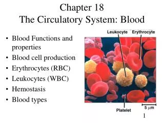
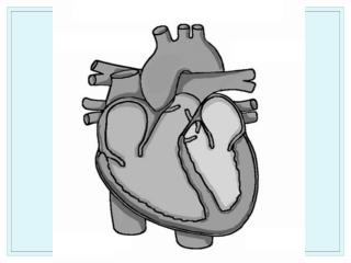
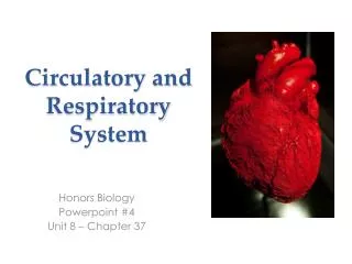
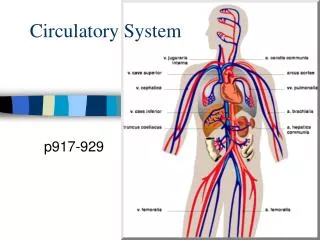
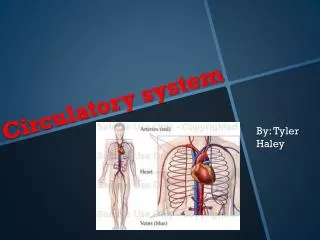
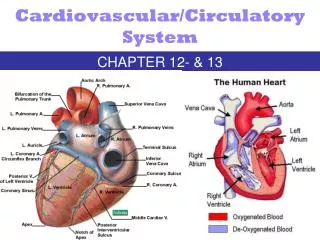
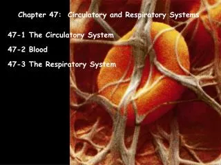
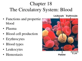
Chapter 18The Circulatory System: Blood • Functions and properties of blood • Plasma • Blood cell production • Erythrocytes • Blood types • Leukocytes • Hemostasis
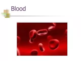
Circulatory System; BloodChapter 18, pg 679 Blood clot showing Red blood cells in a fibrin mesh
Let’s start out with the weird • Drinking blood = strong taboo in most cultures • Except blood sausage & blood pudding both of which are traditional dishes in other countries • It’s one of the rules we kept from the Jewish tradition. • Is there an evolutionary undercurrent that these rules exist to prevent disease transmission? • What about nosebleeds/rare steak? • You can drink a pint of blood before you get sick, says Tyler Durden
The basics, functions and properties • People have 4-6 L of blood • Two components include • Plasma: clear fluid • Cells & Platelets • Erythrocytes (RBCs) • Leukocytes (WBCs) • Centrifuging blood separates the two parts • RBCs make up ~ 45% of volume, a number called the hematocrit • RBCs make blood 4xs as viscous as water
Blood Components • This test tube shows the components of blood in their relative ratios. It shows a hematocrit of 45. The RBC layer together with the "buffy coat" layer make up 45% of the total volume of centrifuged blood (4.5 m. out of 10 ml). • hematocrit of normal adult male : 47 adult female: 42
Plasma • Serum: Like plasma but, without clotting proteins • Proteins of Plasma • Albumins: smallest & most abundant • Regulates osmotic pressure • Globulins: alpha, beta, and gamma • make up antibodies • Fibrogen: allows clotting • Nitrogenous wastes in plasma (urea) are excreted in the kidneys
Erythrocytes (RBCs) • O2 & CO2 carrier • Determine bloodtype • Need to be resilient to get through capillaries • Hemoglobins make up 33% of the cytoplasm • Nucleus is lost during cell formation
Qualities of Erythrocytes • RBC count (Hematocrit) tells how much O2 blood carries • Why women have lower hematocrits • Androgens stimulate RBC production • Menstrual loss • Inverse proportion to body fat • Males also clot faster. • What evolutionary significance might this have?
Erythrocyte Disorders • Polycythemia: Excess RBC • Anemia: RBC Shortage • Sickle Cell: ~1.3 % of African Americans • Symptoms: aches in joints from clogged capillaries, some associated symptoms can be fatal
Malaria • Malaria is caused by parasites that destroy red blood cells. • A symptom is an enlarged spleen, trying to make more RBC’s • Compare distribution area of sickle cell gene with distribution of Malaria
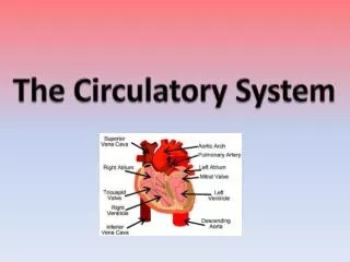
Blood Types • Antigens on RBC surface allow antibodies to recognize what is and what is not us • ABO blood group is a multiple allele explanation of blood types
Blood Compatibility • Agglutination happens when antibodies attack foreign RBCs • AB is called the universal recipient because it has no RBC antibodies • But the donors Antibodies can attack the recipients • Also one of the rarer blood types • O is the universal donor
Rh Groups • Named for Rhesus Monkey • 3 genes, C, D, and E, each with two alleles • DD, or Dd have D antigens on RBCs, • Classified as Rh+ • Rh- lack D antigens • Combined with ABO group to get Blood types like A positive or B negative
Rh Transfusion problems • If Rh- person recieves Rh+ blood • First one is okay, the body hasn’t made any Anti-D antibodies • Second one can cause problems • With fetuses with different Rh groups • The pregnancy is fine as long as there is no tearing of the placenta • Then the baby might be born with Hemolytic disease of the new born (HDN), a type of anemia
Mismatched Transfusion Reaction • Agglutinated RBCs block blood vessels & rupture • free Hb can block kidney tubules & cause death • Universal donors and recipients • AB called universal recipient since it lacks both antibody A and B; O called universal donor • problem is donor’s plasma may have antibodies against recipient’s red blood cells • solution is giving packed cells with minimum plasma
The Rh Group • Rh or D agglutinogens discovered in rhesus monkey in 1940 • blood type is Rh+ if agglutinogens present on RBCs • Rh frequencies vary among ethnic groups • Anti-D agglutinins are not normally present in blood • form only in individuals exposed to Rh+ blood • Rh- pregnant woman carrying an Rh+ fetus or blood transfusion of Rh+ blood • no problems result with either the first transfusion or the first pregnancy, abortion or miscarriage • hemolytic disease of the newborn (erythroblastosis fetalis) occurs if mother has formed antibodies & is pregnant with 2nd Rh+ child • RhoGAM is given to pregnant woman to prevent antibody formation and prevent any future problems • RhoGAM binds fetal agglutinogens in her blood so she will not form antibodies against them during the pregnancy
Hemolytic Disease of Newborn • Mother’s antibodies attack fetal blood causing severe anemia & toxic brain syndrome from excessive bilirubin in blood • treatment is phototherapy to degrade bilirubin or exchange transfusion to completely replace infant’s blood
Other Blood groups • ~100 others, and ~500 antigens • MN, Duffey, Kell, Kidd, and Lewis groups • Rarely cause transfusion problems • Useful in paternity cases
Blood Types • RBC antigens • called agglutinogens A & B • inherited combinations of proteins, glycoproteins and glycolipids on red blood cell • Plasma antibodies • called agglutinins anti-A & -B • gamma globulins in blood plasma that recognize (stick to) foreign agglutinogens on RBCs • responsible for RBC agglutination in mismatched blood transfusions
The ABO Group • Your ABO blood type is determined by presence or absence of antigens (agglutinogens) A & B on RBCs • blood type A person has A antigens, blood type B person has B antigens, AB has both & blood type O has neither • blood type O is the most common; AB the rarest • Antibodies (agglutinins) appear 2-8 months after birth & are at maximum concentration at 10 yr. • antibodies A and/or B, both or none are in plasma • you do not have those that would react against your own antigens • each antibody can attach to several antigens at the same time causing agglutination (clumping)
Hemophilia and European royalty • An X-linked trait, but some get it as a spontaneous mutation • Trouble with clotting factor VIII • The incidence of hemophilia is about 1:7,500 live male births and 1:25,000,000 live female births. Low because we can I.D. it • Transfusions = AIDS trouble
B12 deficiency and anemia • Usually eat 5-7 µgs day. • From meat/milk If you’re not absorbing B12 in your GI tract it can lead to anemia • Like if you have a bleeding ulcer and need part of your stomach removed • Anemia: low RBC count or low hemoglobin
Leukocytes • White blood cells • Have nuclei • Different types are noted by shape of nucleus • Grainy appearance when stained
WBCs Neutrophils • Make up the largest % of WBCs • Releases antimicrobial chemicals • A high count is a sign of bacterial infection Lymphocytes • About 1/3 of WBCs • Fights foreign bodies • Secretes antibodies
Leukemia • Leukemia is cancer of the blood cells. • body produces large numbers of abnormal WBCs • Symptoms • Fever, chills and other flu-like symptoms • Weakness and fatigue • Loss of appetite and/or weight • Swollen or tender lymph nodes, liver or spleen • Easy bleeding or bruising • Tiny red spots (called petechiae) under the skin • Swollen or bleeding gums • Sweating, especially at night • Bone or joint pain • Treatments • Chemotherapy • Radiation therapy • Antibody therapy • Bone Marrow Transplants Also a feline variant
Functions and Properties of Blood • Functions in respiration, nutrition, waste elimination, thermoregulation, immune defense, water and pH balance, etc. • Adults have 4-6 L of blood • plasma, a clear extracellular fluid • formed elements (blood cells and platelets) • Properties of blood • viscosity (resistance to flow) • osmolarity (total molarity of dissolved particles) • if too high, fluid absorption into the blood causes high BP • if too low, fluid remains in the tissues causing edema • one cause is deficiency of plasma protein due to diet or disease
Hematocrit • Centrifuging blood forces formed elements to separate from plasma • Hematocrit is % of total volume that is cells
Plasma and Plasma Proteins • Plasma is a mixture of proteins, enzymes, nutrients, wastes, hormones, and gases • if allowed to clot, what remains is called serum • 3 major categories of plasma proteins • albumins are most abundant plasma protein • contributes to viscosity and osmolarity and influences blood pressure, flow and fluid balance • globulins (antibodies) provide immune system defenses • alpha, beta and gamma globulins • fibrinogen is precursor of fibrin threads that help form blood clots • All plasma proteins formed by liver except globulins (produced by plasma cells descended from B lymphocytes)
Nonprotein Components of Plasma • Plasma contains nitrogenous compounds • amino acids from dietary protein or tissue breakdown • nitrogenous wastes(urea) are toxic end products of catabolism • normally removed from the blood by the kidneys • Nutrients (glucose, vitamins, fats, minerals, etc) • Some O2 and CO2 are transported in plasma • Many electrolytes are found in plasma • sodium makes up 90% of plasma cations accounting for more of blood’s osmolarity than any other solute
Blood Cell Production (Hemopoiesis) • Hemopoietic tissues produce blood cells • yolk sac in vertebrate embryo produce stem cells that colonize fetal bone marrow, liver, spleen & thymus • liver stops producing blood cells at birth, but spleen and thymus remain involved with WBC production • lymphoid hemopoiesis occurs in widely distributed lymphoid tissues (thymus, tonsils, lymph nodes, spleen & peyers patches in intestines) • red bone marrow produces RBCs, WBCs and platelets • stem cells called hemocytoblasts multiply continually & are pluripotent (capable of differentiating into multiple cell lines) • committed cells are destined to continue down one specific cell line • Stimulated by erythropoietin, thrombopoietin & colony-stimulating factors (CSFs)
Erythrocyte Production • Erythropoiesis produces 2.5 million RBCs/second from stem cells (hemocytoblasts) in bone marrow • First committed cell is proerythroblast • has receptors for erythropoietin (EPO) from kidneys • Erythroblasts multiply & synthesize hemoglobin • Normoblasts discard their nucleus to form a reticulocyte • named for fine network of endoplasmic reticulum • enters bloodstream as 0.5 to 1.5% of circulating RBCs • Development takes 3-5 days & involves • reduction in cell size, increase in cell number, synthesis of hemoglobin & loss of nucleus • blood loss speeds up the process increasing reticulocyte count
Erythrocyte Homeostasis • Classic negative feedback control • drop in RBC count causes hypoxemia to kidneys • EPO production • stimulation of bone marrow • RBC count in 3-4 days • Stimulus for erythropoiesis • low levels of atmospheric O2 • increase in exercise • hemorrhaging
Nutritional Needs for Erythropoiesis • Iron is key nutritional requirement for erythropoiesis • lost daily through urine, feces, and bleeding • men 0.9 mg/day and women 1.7 mg/day • low absorption rate requires consumption of 5-20 mg/day • dietary iron in 2 forms: ferric (Fe+3) & ferrous (Fe+2) • stomach acid converts Fe+3 to absorbable Fe+2 • gastroferritin from stomach binds Fe+2 & transports it to intestine • absorbed into blood & binds to transferrin to travel • bone marrow uses to make hemoglobin, muscle used to make myoglobin and all cells use to make cytochromes in mitochondria • liver binds surplus to apoferritin to create ferritin for storage • B12 & folic acid (for rapid cell division) and C & copper for cofactors for enzymes synthesizing RBCs
Leukocyte Production (Leukopoiesis) • Committed cell types -- B & T progenitors and granulocyte-macrophage colony-forming units • possess receptors for colony-stimulating factors • released by mature WBCs in response to infections • RBC stores & releases granulocytes & monocytes • Some lymphocytes leave bone marrow unfinished • go to thymus to complete their development (T cells) • Circulating WBCs do not stay in bloodstream • granulocytes leave in 8 hours & live 5 days longer • monocytes leave in 20 hours, transform into macrophages and live for several years • WBCs providing long-term immunity last decades
Platelet Production (Thrombopoiesis) • Hemocytoblast that develops receptors for thrombopoietin from liver or kidney becomes megakaryoblast • Megakaryoblast repeatedly replicates its DNA without dividing • forms gigantic cell called megakaryocyte (100 m in diameter that remains in bone marrow) • Infoldings of megakaryocyte cytoplasm splits off cell fragments that enter the bloodstream as platelets (live for 10 days) • some stored in spleen & released as needed
Erythrocytes (RBCs) • Disc-shaped cell with thick rim • 7.5 M diameter & 2.0 m thick at rim • Major function is gas transport • lost all organelles during maturation so has increased surface area/volume ratio • increases diffusion rate of substances in & out of cell • 33% of cytoplasm is hemoglobin (Hb) • O2 delivery to tissue and CO2 transport back to lungs • contains enzyme, carbonic anhydrase (CAH) • produces carbonic acid from CO2 and water • important role in gas transport & pH balance
Hemoglobin Structure • Hemoglobin consists of 4 protein chains called globins (2 alpha & 2 beta) • Each protein chain is conjugated with a heme group which binds oxygen to ferrous ion (Fe+2) • Hemoglobin molecule can carry four O2 • Fetal hemoglobin has gamma instead of beta chains
Erythrocytes and Hemoglobin • RBC count & hemoglobin concentration indicate the amount of oxygen the blood can carry • hematocrit(packed cell volume) is % of blood composed of cells • men 42-52% cells; women 37-48% cells • hemoglobin concentration of whole blood • men 13-18g/dL; women 12-16g/dL • RBC count • men 4.6-6.2 million/L; women 4-2-5.4 million/L • Values are lower in women • androgens stimulate RBC production • women have periodic menstrual losses
Erythrocyte Death & Disposal • RBCs live for 120 days • membrane fragility -- lysis in narrow channels in the spleen • Macrophages in spleen • digest membrane bits • separate heme from globin • hydrolyze globin (amino acids) • remove iron from heme • convert heme to biliverdin • convert biliverdin to bilirubin • becomes bile product in feces
Erythrocyte Disorders • Polycythemia is an excess of RBC • primary polycythemia is due to cancer of erythropoietic cell line in the red bone marrow • RBC count as high as 11 million/L; hematocrit of 80% • secondary polycythemia from dehydration, emphysema, high altitude, or physical conditioning • RBC count only up to 8 million/L • Dangers of polycythemia • increased blood volume, pressure and viscosity can lead to embolism, stroke or heart failure
Anemia - Deficiency of RBCs or Hb • Causes of anemia • inadequate erythropoiesis or hemoglobin synthesis • inadequate vitamin B12 from poor nutrition or lack of intrinsic factor from glands of the stomach (pernicious anemia) • iron-deficiency anemia • kidney failure & insufficient erythropoietin hormone • aplastic anemia is complete cessation (cause unknown) • hemorrhagic anemias from loss of blood • hemolytic anemias from RBC destruction • Effects of anemia • tissue hypoxia and necrosis (short of breath & lethargic) • low blood osmolarity (tissue edema) • low blood viscosity (heart races & pressure drops)