Download
1 / 1
10 likes | 117 Vues
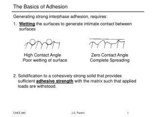
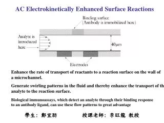
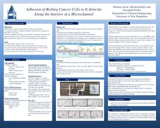
Flow Direction. Brittany Artale , Michael Balch, and Alexandra Eicher Department of Chemical Engineering University of New Hampshire. Adhesion of Rolling Cancer Cells to E- Selectin Along the Interior of a Microchannel. A: No flow. B: Start of flow. C: Clump of cells stop moving.

E N D
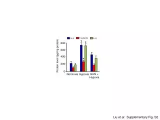
Flow Direction Brittany Artale, Michael Balch, and Alexandra EicherDepartment of Chemical EngineeringUniversity of New Hampshire Adhesion of Rolling Cancer Cells to E-Selectin Along the Interior of a Microchannel A: No flow. B: Start of flow. C: Clump of cells stop moving. Motivation & Goals Introduction Results & Discussion • Motivation: • Metastasis is of great concern when treating cancer patients • Localized radiation or surgical treatments will not destroy migrated or metastatic cancer cells • It may be possible to develop a microfluidic system that can selectively remove metastatic cancer cells from a blood steam, using a selective adhesion molecule E-selectin, to prevent further tumor establishment • Goals: • To successfully culture a strain of cancer cells • To fabricate a microfluidic system to study the flow of cancer cells • To coat a section of some of the microfluidic devices with E-selectin • To flow cancer cells through these treated channels • To observe the cancer cells’ adhesion properties to E-selectin • Cancer Cells: • Abnormal cells that no longer express the basic characteristics of healthy cells • Propagated from multiple mammalian cell mutations • A major property of cancer cells is their ability to metastasize • Metastasis is the ability of a cell to spread to other locations in the body • Metastasis is accomplished by circulating cancer cell adhesion to the wall of a blood vessel, its penetration through this wall, and its formation into a tumor in the surrounding tissue (See Figure 1) • E-selectin: • Prominent cell adhesion molecule found on the surface of endothelial cells • Carbohydrates on the surface of cells can easily adhere to E-selectin (See Figure 2) • Carbohydrates are increased on the surface of cancer cells • Cancer cells seem more suited than healthy cells to adhere to endothelial cells at metastatic sites • Based on the various methods to construct the microfluidic devices ,the use of household glue to create a straight-channel mold provided the cleanest cast. These are the optimum techniques that should be used when creating new casts. • Plasma cleaning was successful in adhering the cast to the PDMS coated and glass slides. The microfluidic devices were successfully fabricated to study the adhesion properties of E-selectin to cancer cells in solution. One of the microfluidic devices is shown in Figure 3. • The channels that were treated showed evidence of cell adhesion, while the control channels did not. Figure 4 shows one observation of a cell rolling and adhering to the surface in the treated channel. Figure 5 shows other cells that are still flowing through the device, providing evidence of adhesion. This adhesion to the channel walls was not observed in the untreated channel. Figure 2: Depiction of Cellular Adhesion to E-selectin1 Figure 1: Depiction of Metastasis1 Methods Conclusion • Cell Culturing: • Cell lines cultured: • LS174T - human colon carcinoma • MCF7 - human breast carcinoma • Cultured using standard cell culturing techniques: • CO2 independent medium (1X) • 10% Fetal Bovine Serum • 2 mM L-Glutamine • Device Mold Preparation: • Adhered 27 gauge needles to glass slides: • Elmer’s glue • House glue • Channel Preparation: • 10:1 ratio of PolyDimethylsiloxane (PDMS) to curing agent • Adhered casts to slides: • PDMS coated • Glass • Control Channel Treatment: • Flowed milk solution through the entire channel system to prevent cells adhering to PDMS channel walls • Washed channels with PBS • Channel Treatment: • Inserted a slug of 1.11 µg/mL Protein G in PBS into each channel • Washed Channels with PBS • Inserted a slug of 2 µg/mL E-selectin in PBS over the treated area • Second wash with PBS • Flowed milk solution through the entire channel system, and let sit for 30 minutes • Washed channels with 0.1 M Ca+ Mg+ in PBS to activate E-selectin • Flow Tests: • Set flow rate between 5 µL/min and 10 µL/min of a PBS solution with a cell concentration of 104 cells/mL to flow through the treated and control devices • Observed channels with an inverted microscope and camera for half an hour • Successfully fabricated channels • Determined optimum techniques for channel fabrication • Glue type – household glue • Geometry – straight-channel • Observed cellular adherence to the walls of treated channels Future Work • Quantify the differences between treated and untreated channels by recording the number of cells that became adhered to the respective microchannelwalls • Determine bond strength of E-selectin by increasing the flow rate of the PBS solution until adhered cells and flushed away • Use the required flow rate to flush away adhered cells to calculate the shear stress and in turn force • Fabricate new molds and channel systems using the optimum techniques • Explore shelf life of E-selectin treatment Data Figure 3: Photograph of one of our microfluidic devices. This one was made using house glue and a PDMS coated glass slide. References A C B http://www.bio.davidson.edu/courses/immunology/students/spring2006/latting/home%20copy.html Figure 4: Depiction of a clump of cancer cells sticking to the wall of a treated microfluidic device. Flow Direction Flow Direction Flow Direction Flow Direction Flow Direction Flow Direction Acknowledgments We would like to thank Professor Adam St. Jean for mentoring us through this project, Professor Russell Carr for showing us the techniques needed to created the microfluidic devices, and Professor Nivedita Gupta for allowing us to use her laboratory resources. D A B C Figure 5: Depiction of a clump of cancer cells stuck to the wall of a treated microfluidic device.