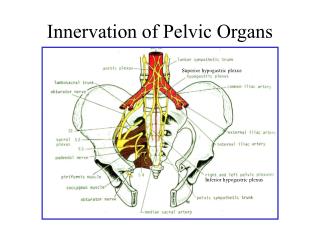
Anatomy of the Female Pelvic Organs
830 likes | 984 Vues
Explore the anatomy of female pelvic organs, including the vulva, vagina, cervix, and uterus. Learn about structures, functions, and relationships for a comprehensive understanding. Essential for medical professionals and students.

Anatomy of the Female Pelvic Organs
E N D
Presentation Transcript
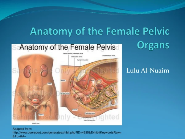
Anatomy of the Female Pelvic Organs Lulu Al-Nuaim Adapted from: http://www.doereport.com/generateexhibit.php?ID=4935&ExhibitKeywordsRaw=&TL=&A=
Aims • To fully understand the anatomy of the female pelvis in terms of bones and tissues, and fetal skull, this would help in explaining the mechanism of Labour & Delivery. AL Nuaim
Objectives • Student at the end of session should be able to: • Describe anatomy of female bonny pelvis&Diameters. • Discuss the important landmarks in the female pelvis. • To know the types of pelvis. • Comprehend the normal organs with their blood, venous, lymphatic drainage and innervation. • Explain the relationship between pelvic organs. • Understand the relationship between the female pelvis (Bones& Soft Tissue) and fetal skull, in order to understand the mechanism of labour AL Nuaim
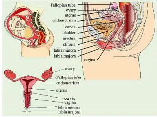
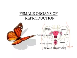
The Vulva external organs of the female Include: - • Mons veneris • Labia majora • Labia minora • The clitoris • The vestibule The intriotus has six openings: • Urethral meatus • Two skene’s ducts • Vaginal orifice • Two Bartholin ducts. Adapted from: https://www.cancer.org/cancer/vulvar-cancer/about/what-is-vulvar-cancer.html AL Nuaim
The Vulva Bartholin glands - lies on each side of the vagina, in the posterior lower third 1/3 of the interiotus. • Secrete mucus – alkaline • Blood supply: Pudendal artery from the femoral aa Venous drainage in the corresponding vein. • Lymphytic inguinal glands External iliac glands • Nerves: Branches of the pudendal nerve, perineal nerve (T12 L1-2, S2-4) • In labour: Catheterization, Episiotomy, Anaesthetic infiltration AL Nuaim
The vagina A Canal/tube extend from the vulva to the uterus • Runs upwards and backwards • Walls lie in close contact, easily separated. Speculum examination • The posterior vaginal wall is longer than the anterior 11.5 cm (4.5 in) vs 7.5 cm • Cervix enters the vagina at a right angle. • Tornices = four Anterior, posterior and Two laterals AL Nuaim
Adapted: https://img.webmd.com/dtmcms/live/webmd/consumer_assets/site_images/articles/image_article_collections/anatomy_pages/vagina.jpg?resize=646px:*&output-quality=100 AL Nuaim
The Vagina Blood supply • Vaginal aa, uterine aa, middle haemorridal, inferior vesical, pudendal branches of the internal iliac aa. • Venous drainage to corresponding veins. • Lymph: inguinal, internal iliac, sacral glands • Nerves: symphatetic and parasymphatetic • Relations: Anterior : base of the bladder on upper ½ of vagina. cystocele Posterior: upper ½ ???Pouch of Douglas in the lower ½ Rectocele Rectum centrally Perineal body inferiorly Laterally: AL Nuaim
The Cervix Forms the lower 1/3 of the uterus • Enter the vagina at a right angle • Barrel shape • 2.5 cm (1 in) long • Two parts: • Supravaginal • Intra vaginal • Cervical os • Internal os • External os; shape differ in nulliparous and multiparous • Cervical canal between the internal os and the external os • Cervical canal is covered by columnar epithelium • Transformation zone; squamous-columnar junction. • Cervical Ectropion AL Nuaim
Adapted from: http://www.medindia.net/images/common/patientinfo/950_400/anatomy-of-the-cervix.jpg AL Nuaim
Blood supply Uterine aa Lymphatic drainage Internal iliac, sacral glands Adapted from: https://www.earthslab.com/wp-content/uploads/2017/03/031717_1945_Uterus8.png AL Nuaim
The Cervix Supports: • Cardinal ligaments/Transverse Cervical • Pubocervical ligaments • Uterosacral ligaments All 3 ligaments insert into supra-vaginal cervix &upper vagina In pregnancy: • Rich blood supply – bluish coloration • Soft • Cervical glands – mucus plug “operculum” Late in pregnancy – softer and starts to dilate. In labor: • The longitudinal smooth muscle fibres of the uterus contract and retract pulling upward thus reducing the length of the cervix. Cervical Bishop Score • The cervix is made up of fibrous and elastic tissue • Full dilatation marks the end of the first stage of labour. AL Nuaim
The Uterus The uterus lies in the true pelvis. Anteverted (A/V)and anteflexed (A/F)in position. The body of the uterus lies above the bladder. • Size: 7.5 cm length • 5 cm wide • 2.5 cm thick • 50 -75 gm weight, in pregnancy 1 Kg ???? • Position/site: Gross structure: • The cervix lower 1/3 • The isthmus • The cavity • The corpus • The cornua. • The fundus AL Nuaim
Adapted from: http://aibolita.com/uploads/posts/2015-03/44qv-132.jpg AL Nuaim
Layers: Endometrium Myometrium Perimetrium - peritoneum Adherent, where??? Loose,??? Blood supply: Arteries: fundus – ovarian artery (aa) Body-uterine aa , directly from internal iliac aa Venous: Rt Ovarian???? Left Ovarian??? The relationship between the ureter and uterine aa • Uterine aa runs behind the peritonieum, cross transeverse cervical ligament (Cardinal ligament) then the aa passes anterior to and above the Ureter 1.5cm from lateral vaginal wall fornix. Water under the bridge, Important landmark AL Nuaim
Venous: Right ovarian vein - inferior vena cava Left ovarian vein – renal vein Lymph Internal and external iliac glands Inguinal /Sacral glands Nerves: Sympathatic and Parasympathetic AL Nuaim
THE FALLOPIAN TUBEs • Extend from the cornua of the uterus, travels towards the sidewalls of the pelvis. Then turns downwards and backwards. • The tube lies in the upper margin of the broad ligaments • Communicate; superiorly with the uterine cavity, Inferiorly with the perineal cavity. Tubal patency checked by different means • Length 10 cm : 3 mm thick • 4 PARTS • Interstitial • Ampula • Infundebulum • Fimbrial • BLOOD SUPPLY - ovarian aa Uterine aa Venous drainage by corresponding veins AL Nuaim
THE OVARIES • Lie in the posterior wall of the broad ligament at the fibrial end of the fallopian tubes at the level of the pelvic brim. • Size: almond like = 3 x 2 x 1.5 cm Dull white colour, Corrugated surface • Structure varies with woman’s age. AL Nuaim
The Ovaries • Blood supply – ovarian aa • Ovarian vein • Lymphatic lumbar glands • Nerves ovarian plexus • SUPPORTS They lie in a fossa, posterior leafof broad ligament • Attached to broad ligament – meso ovarian • The meso salpinx is the broad ligament that extend between the fallopian tube and the ovary. • The Fallopian tubes, ovaries and broad ligaments are called Adenxa Bimanual examination AL Nuaim
Adapted from: https://download.e-bookshelf.de/download/0000/5952/91/L-X-0000595291-0001346903.XHTML/images/c01f004.jpg AL Nuaim
Ligaments: • Round ligaments Maintains uterus in A/V + A/F From the cornua of the uterus – pass downwards and insert in the tissue of the labia majora. • Broad ligaments Not a true ligament Folds of peritoneum extend laterally from the uterus to the pelvic side walls. • Cardinal ligaments • Pubocervical • Uterosacral AL Nuaim
Adapted from: http://ueu.co/wp-content/uploads/2014/09/loadBinaryCASJBB06.jpg AL Nuaim
Adapted from: http://www.integraltheory.org/diagrams/5.jpg AL Nuaim
THE PELVIC FLOOR • The outlet of the pelvis is filled with soft tissue that supports the pelvic and abdominal organs. • It forms as a gutter-shaped structure higher anteriorly than posteriorly. • Three canals, each with an external orifices, run through the tissue are the: • Urethra • Vagina • Rectum AL Nuaim
Adapted from: http://www.beyondbasicsphysicaltherapy.com/images/Stein-Illustration2.jpg AL Nuaim
Pelvic Floor • There are six layers of tissue. • An outer covering of skin • Subcutaneous fat • Superficial muscles enclosed in fascia • Deep muscles enclosed in fascia • Pelvic fascia thickened to form pelvic ligaments • Peritoneum AL Nuaim
Pelvic Floor • Superficial Pelvic Floor muscles: • One muscle Transverse perinei • Two muscles Bulbo-cavernosus • One muscle Ischio-cavernosus The membranous Sphincter of the urethhra and the rectal sphincter • Deep Pelvic Floor muscles Three pairs of muscles all have their insertion around the coccyx, sometimes called cocygeous muscles. Their anatomical name is “levatorani” muscles, 5 mm thick • Iliococcygeus • ischiococcygeus • pubo- coccygeus AL Nuaim
Adapted from: http://obgyn.mhmedical.com/data/books/1307/rog001_fig_02-05.png AL Nuaim
Adapted from: https://image.slidesharecdn.com/gver8awqh2g0vl4z4oc7-signature-e7df6fed128681beaed0608e176de70c6dbea69a6285127762e6c672f362a953-poli-170209224138/95/anatomy-of-pelvis-perineum-25-638.jpg?cb=1486681764 AL Nuaim
The perinuem • Bounded by Levatorani above and the anus below • Divided into: urogenital triangle anteriorly and anal sphincter posteriorly • Covered by superficial and deep fascia AL Nuaim
perineal body • Fibromuscular mass • Lies between the vaginal and rectal canals • Is triangular, the base is the skin and the apex pointing upward each side is 3.8 cm in length • Three layers of tissue 1. outer covering of skin 2. superficial pelvic floor • bubo-cavernous muscle • Ischio-cavernouses muscle • transverse perinei muscle 3. deep pelvic floor muscle. Episiotomy, types, indications, AL Nuaim
THE NORMAL FEMALE PELVIS The pelvis articulate with the fifth lumbar vertebra above and with the head of each femur in the right and left acetabulum. • The weight of the trunk is transmitted through the pelvis into the legs. • Gives protection to the pelvic organs • The pelvis is the largest bone in the body. Gross structure: Consists of 4 bones: • 5 fused sacral vertebrae and coccyx • left & right innominate bones AL Nuaim
Adapted from: http://www.eorif.com/Pelvis/Xray%20Pelvis.html AL Nuaim
Adapted from: http://www.open.edu/openlearncreate/pluginfile.php/4415/mod_oucontent/oucontent/14/none/none/fig3.jpg AL Nuaim
The Sacrum A triangular shape; 5 fused vertebrae and 4 pairs of holes (nerves, blood vessels/lymph) The hollow of the sacrum – smooth and concave The alae of the sacrum - give the appearance of wings • The sacral promontary is the centre point of the upper border of the first sacral vertebrae. • The sacral canal opens at the level of 5th sacral vertebra, a passage for spinal cord. • At the level of the 2nd and 3rd sacral vertebrae, the nerves spread out to form the cauda equina. • Anaesthesia&Analgesia in labour AL Nuaim
THE COCCYX • 4 Fusesd coccygeal vertebrae • Triangular shape • Articulate with the sacrum • Muscles are attached to its tip. • Easley in labour AL Nuaim
Right &Left In-nominate Bones Each made of 3 separate parts that meet in the acetabulum. • Ilium upper part is iliac crest (anterior and posterior, superior iliac crest • Ischiumischialtuberosity , 2 cm above is the ischial spines. • Pubis both meet the pubic body fused by cartilage “symphysis pubis” • PELVIC JOINTS • The two sacroiliac joints • The symphysis pubis • The sacrococcygeal joints • THE PELVIC LIGAMENTS • Sacroiliac ligament = strongest in the body • Sacro tuberous • Sacrospinous • Inguinal ligament AL Nuaim
Adapted from: http://www.learnbones.com/wp-content/uploads/Pelvic_girdle.jpg AL Nuaim
DIVISIONS OF THE PELVIS The brim divides the pelvis into two parts: • The false: lies above the pelvic brim not important in obstetrics • The true: what lies below the pelvic brim. It has a : brim , cavity and an outlet Forms the curved canal through which the fetus pass during labour. AL Nuaim
Adapted from: http://teachmeanatomy.info/wp-content/uploads/Greater-and-Lesser-Pelvis-Divided-by-the-Pelvic-Brim.jpg AL Nuaim
The brim or inlet Round in shape; partlybone & partly ligaments • Has eight points as demonstrated • Bounded anteriorly by the pubis • Laterally by illiopectineal lines • Posteriorly by ale and sacral promontory • Widest diameter is, Transverse AL Nuaim
True Conjugate ( Anteroposterior diameter) from sacral promontory to upper inner border of Symphysis pubis, Measured by erect lateral pelvimetry an X-Ray • Obstetric Conjugate From the inner surface of symphysis pubis to the sacral promontory • Diagonal Conjugate, from the sacral promontory to ? AL Nuaim
Adapted from: https://image.slidesharecdn.com/normallabour-140428082740-phpapp02/95/normal-labour-7-638.jpg?cb=1398675297 AL Nuaim
The Pelvic cavity • Extends from the brim above to the pelvic outlet below • The posterior wall 11 cm formed by hollow of the sacrum • The anterior wall is formed by the symphysis pubis and obturator foramen 3.8 cm • The lateral walls sacrosciatic ligamnet and ischial spines • Interspines Diameter AL Nuaim
The pelvic outlet • Anatomical outlet • Obstetrical outlet • The anatomical outlet is formed by fixed pointes useful landmarks for taking pelvic measurement. • Bounded anteriorly by pubic Arch • Laterally by sacrosciaticlig&IschailTuberosity • Posteriorly by tip of Coccyx • The obstetrical outlet The landmarks are: • The lower border of the symphysis pubis • The ischial spines • The sacro-spinous ligament • The lower border of the sacrum. AL Nuaim
Adapted from: http://slideplayer.com/slide/2790499/10/images/7/PELVIC+INLET+PELVIC+OUTLET.jpg AL Nuaim
Average measurements of pelvis • Brim Antero-posterior = 11.5 cm Transverse = 13.0 cm • Cavity Antero-posterior = 12.0 cm Transverse (I/S) = 12.0 cm Outlet Antero-posterior = 12.5 cm Transverse = 11.0 cm AL Nuaim
Abnormal Pelvis Four Types • Gynecoid Pelvis 50% • Anthropoid 25% • Android Pelvis 20% • Platypelloid (flat 5% AL Nuaim