Understanding the Human Heart: Structure, Function, and Circulation
This comprehensive overview covers the anatomy and physiology of the human heart, detailing its four chambers, associated blood vessels, and the pathway of blood circulation through both pulmonary and systemic circuits. It explains how coronary arteries supply oxygen and nutrients, the process of collecting and pumping blood, as well as the dynamic action of heart valves. Additionally, it outlines the control of heartbeat through myogenic contractions, the role of the sinoatrial node, and the nervous system's influence, including adrenaline's effect on heart rate.

Understanding the Human Heart: Structure, Function, and Circulation
E N D
Presentation Transcript
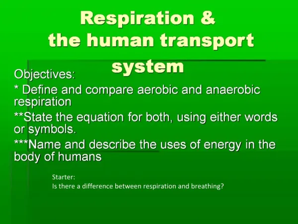
The transport system Topic 6.2
Assessment Statements 6.2.1 Draw and label a diagram of the heart showing the four chambers, associated blood vessels, valves and the route of blood through the heart. 6.2.2 State that the coronary arteries supply heart muscle with oxygen and nutrients. 6.2.3 Explain the action of the heart in terms of collecting blood, pumping blood, and opening and closing of valves. 6.2.4 Outline the control of the heartbeat in terms of myogenic muscle contraction, the role of the pacemaker, nerves, the medulla of the brain and epinephrine (adrenaline). 6.2.5 Explain the relationship between the structure and function of arteries, capillaries and veins. 6.2.6 State that blood is composed of plasma, erythrocytes, leucocytes (phagocytes and lymphocytes) and platelets. 6.2.7 State that the following are transported by the blood: nutrients, oxygen, carbon dioxide, hormones, antibodies, urea and heat.
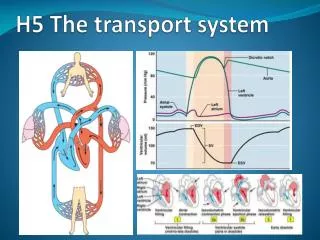
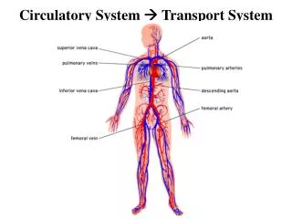
The human heart • Pair of side-by-side pumps • Two atria • Thin-walled collection chambers • Two ventricles • Thick-walled muscular pump • Produces force known as blood pressure • Blood moves through large arteries, smaller arteries, arteriole, capillary bed, venule, larger vein, largest veins
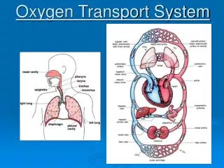
Two circuits • Pulmonary circuit • Right side of heart sends blood to lungs • Systemic circuit • Left side of heart sends blood to body cells
Pulmonary circulation • Deoxygenated blood comes from body • Enters heart through vena cava • Blood collects in right atrium (RA) • Moves through rt. atrioventricular (AV) valve to rt. ventricle • RA contracts forcing any remaining blood into the right ventricle (RV) • Once a volume of blood has accumulated in the RV, it begins to contract
Rt. AV valve closes to prevent backflow to the RA (closing of valve creates ‘lub dub’ sounds heard through a stethoscope) • Dramatic increase in blood pressure (BP) inside the RV opens the right semilunar valve • Blood enters pulmonary artery (PA) • Blood leaves PA and enters lungs through smaller arteries and capillaries • RBCs drop off carbon dioxide and pick up oxygen
Systemic circulation • RBC enters left atrium (LA) • Blood accumulates in LA • Blood travels through left AV valve and enters the left ventricle (LV) • LV contracts • Left AV valve closes to prevent backflow into the LA • Dramatic increase in bp inside the LV opens the left semilunar valve • Blood enters the aorta and then to body
Coronary arteries • First arteries to receive blood that has left the heart • Coronary arteries branch into the heart muscle itself • Supply the heart with oxygen and nutrients
Control of heart rate • The majority of tissue making up the heart is cardiac muscle • It contracts and relaxes without nervous system control (known as myogenic muscle contraction) • Needs to be controlled in order to keep the timing of the contractions unified and useful • Right atrium conatins a mass of tissue within its walls known as the sinoatrial node (SA node)
Sinoatrial Node • Mass of tissue acts as the pacemaker for the heart • Sends out an ‘electrical’ signal to initiate the contraction for both atria • Resting heart rate of 72 bpm, signal from the SA node is sent out every 0.8 seconds
Atrioventricular node • Also within the right atrium • AV node receives the signal from the SA node, waits approximately 0.1 seconds and then sends out another ‘electrical’ signal • Signal goes to the ventricles and results in their contraction
Medulla • During times of increased body activity, the heart rate needs to increase to get rid of excess CO2 and gain more O2 • As CO2 levels rise, an area of the brainstem called the medulla chemically senses the increase • Signal sent through the cardiac nerve to the SA node to increase heart rate to an appropriate level • Timing is changed
After exercise • Level of CO2 in bloodstream begins to decrease and another signal is sent from the medulla by means of the vagus nerve • the SA node once again takes over the timing of the heart rate
Other influences • During periods of high stress or excitement, adrenal glands secrete adrenaline into the bloodstream • Causes SA node to ‘fire’ more frequently than it does at the resting heart rate and thus heart rate increases
Arteries • Arteries are blood vessels taking blood away from the heart that has not yet reached a capillary • Have thick smooth muscle layer that is used by your autonomic nervous system to change the inside diameter of the blood vessels which helps to regulate bp • Arteries→arterioles→capillary bed→venule
Capillaries • Blood enters a capillary bed, much of the pressure is lost • blood cells make their way through one at a time • Chemical exchanges occur through the single-cell thickness of capillaries
Veins • Veins receive blood at a relatively low pressure from the capillary beds • Blood flow is slower • Collect blood from capillaries and return it the heart • Veins have thin walls and a larger internal diameter • Have many internal passive valves that help keep the slow-moving blood consistently moving towards the heart