6.2 The Transport System
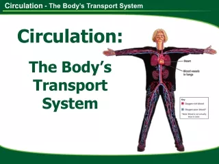
6.2 The Transport System. Our circulatory system provides a delivery and collection service for the whole body. The heart, blood and blood vessels make up an efficient transport system that reaches all cells, bringing the substances they need and taking away wastes.

6.2 The Transport System
E N D
Presentation Transcript
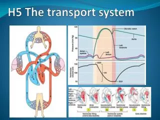
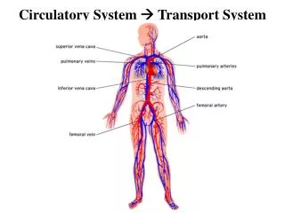
Our circulatory system provides a delivery and collection service for the whole body. The heart, blood and blood vessels make up an efficient transport system that reaches all cells, bringing the substances they need and taking away wastes. Humans and other mammals have a closed circulatory system with blood contained inside a network of arteries, veins and capillaries
The Heart Blood is kept moving by the pumping action of the heart muscle. The heart will beat 2.5x10⁹ times in a lifetime on average, sending more than 1.5 million liters of blood from each ventricle. The heart is about the size of your fist. It is a double pump with two separate sides. The right hand receives deoxygenated blood from all over the body and pumps it to the lungs to pick up more oxygen. The left hand side receives oxygenated blood from the lungs and pumps it to cells all over the body.
The heart has four chambers- two smaller atria (atrium singular) at the top. Two larger ventricles below. The right and left hand sides are completely separated from one another. Atria have thin walls as the blood received from the veins is under relatively low pressure. Ventricles are stronger and more muscular as their job is to pump blood out of the heart. Both ventricles hold the same volume of blood but the left ventricle is thicker than the right since it must generate enough pressure to pump blood all around the body. The right pumps only to the lungs. http://www.youtube.com/watch?v=DAXa4eR1s0M http://www.youtube.com/watch?v=oE8tGkP5_tc
Atria are separated from ventricles by atrioventricular valves, which prevent the blood flowing backwards into the atria. A second set of valves in the aorta and pulmonary arteries- the semilunar valves- prevent backflow into the ventricles as they relax after contraction. Heart muscle works continuously, beating 75 times per minute when an average person is resting. Coronary arteries extend over the surface of the heart and penetrate deep into the muscle fibers to supply oxygen and nutrients.
The Cardiac Cycle The cardiac cycle is the sequence of events that takes places during one heart beat. As the heart’s chambers contract, blood inside them is forced on its way. Valves in the heart and arteries stop the blood flowing backwards.
1. 4. 3. 2 Atrial Systole- The muscles of the atrium wall contract, pushing blood through the atrioventricular valves in the ventricles. Both atria contract at the same time. Ventricular Systole- Blood forced into the ventricles causes blood pressure inside to rise, so the antroventricular valves snap closed. When the ventricles are full, ventricle muscles contract, generating the pressure that drives blood through the semilunar valves in the aorta and pulmonary artery. A pulse is produced that can be felt in arteries in other parts of the body.
1. 4. 3. http://www.youtube.com/watch?v=rguztY8aqpk 2 3. Diastole- Ventricles and atria now relax, and the pressure inside them is low. The semilunar valves are closed by the back pressure of blood in the arteries. 4. Blood flows into the atria from the veins, opens the atrioventricular valves, and begins to fill the ventricles. Blood from the body enters the right atrium via the vena cava. Blood from the lungs enters the left atrium from the pulmonary vein. 5. The whole cycle repeats when the atria contract again.
Control of the Heart Beat Heart tissue is made of a special type of muscle that is different from other muscles in our bodies. Cardiac muscle is unique because it contracts and relaxes without stimulation from the nervous system. It is said to be myogenic. Natural myogenic contractions are initiated at an inbuilt pacemaker, which keeps cardiac muscle working in a coordinated, controlled sequence. The pacemaker, or sinoatrial node (SAN), is a special region of muscle cells in the right atrium that sets the basic pace of the heart. The rate set by the SAN is also influenced by stimulation from the nervous system and by hormones.
At the start of every heart beat, the SAN produces an impulse that stimulates both atria to contract. A second structure, the atrioventricular node (AVN) at the base of the right atrium, is also stimulated. It delays the impulse briefly until the atrila contraction finishes and then transmits it on a bundle of modified muscle fibers to the base of the ventricles. Impulses radiate up through the ventricles, about 0.1 seconds after the atria.
The natural rhythm of the pacemaker is modulated by the nervous system so that the heart rate is adjusted to our activity levels. It speeds up when exercising and more oxygen and nutrients are needed, and slows down when we sleep. Changes to our heart rate are not under our conscious control but result from impulses sent from a control center in the part of the brain stem known as the medulla. http://www.youtube.com/watch?v=yGlFBzaTuoI
Impulses to speed up the heart pass along the sympathetic nerve, which stimulates the pacemaker to increase rate. Impulses sent along he parasympathetic nerve cause the heart rate to slow down. The medulla monitors blood pressure and CO2 levels. Emotions such as stress, as well as increases in activity level, can cause increase in heart rate. During periods of excitement, hear or stress the adrenal glands release the hormone adrenalin, which travels in the blood to the pacemaker and stimulates it to increase the heart rate.
Blood and Blood Vessels Endothelium- very smooth single layer of cells Lumen Collagen fibers Arteries are blood vessels that carry blood away from the heart. They branch and divide many times before forming arterioles and eventually tiny capillaries that reach all the tissue in the body. Arteries have thick outer walls of collagen and elastic fibers which withstand high blood pressure and prevent vessels becoming overstretched or bursting. Just beneath the outer covering is a ring of circular smooth muscle that contracts with each heart beat to maintain blood pressure and keep blood moving. Inside the artery, the lumen is narrow to keep blood pressure high. The lumen’s lining of smooth epithelial cells reduces friction and keeps blood moving. Elastic fibers and smooth muscle
Capillaries Capillaries are the smallest vessels The lumen of the capillary is only about 10um in diameter and some are so small that red blood cells must bold up in order to pass along. Networks of these tiny capillaries reach almost every body cell. Blood flow here is very slow, at less than 1mm per second, but capillary walls are only one cell thick so the distance for diffusion of materials in and out of them is as small as possible. Some capillaries have spaces between their cells enabling plasma and phagocytes to leak out into the tissues.
Veins Veins carry blood back towards the heart from body tissues. Small veins called venules join up to form large veins, which have much thinner walls than arteries. They also contain few elastic and muscle fibers. Blood inside a vein does not pulse along and the lumen is large to hold slow-moving blood. The thin walls can be compressed by adjacent muscles and this helps squeeze blood along and keep it moving. Many veins contain valves to prevent blood from flowing backwards.
Composition of Blood Blood plasma is a pale yellow liquid that makes up 50-60% of blood volume. Suspended in plasma are three important groups of cells: Eryhrocytes (red blood cells), whose job is to carry oxygen Leucocytes (white blood cells), which fight disease. Platelets (cell fragments), which are needed for blood clotting
Functions of Blood Blood has two important roles: Carrying dissolved materials to all cells, Helps fight infectious diseases.