Download
1 / 1
20 likes | 177 Vues
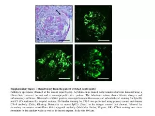
a. b. Supplementary figure 1 : Renal biopsy from the patient with IgA nephropathy

E N D
a b Supplementary figure 1: Renal biopsy from the patient with IgA nephropathy Pathology specimens obtained at the second renal biopsy. A) Glomerulus stained with hematoxylin/eosin demonstrating a fibrocellular crescent (arrow) and a mesangioproliferative pattern. The tubulointerstitium shows fibrotic changes and inflammatory infiltrates. Glomeruli exhibited positive mesangialimmunofluorescent and subendothelial staining for IgA (B) and C3 (C) performed by hospital routines. D) Similar staining for C5b-9 was performed using primary mouse anti-human C5b-9 antibody (Dako, Glostrup, Denmark), or mouse IgG2a (Dako) as the isotype control (not shown), followed by secondary anti-mouse Alexa-Fluor 488-conjugated antibody (Molecular Probes, Eugene, OR). C5b-9 staining was more prominent in the capillary walls as well as in the mesangium. Scale bars 100 μm. d c