Download
1 / 28
290 likes | 521 Vues
Lymphocyte Activation Recognition David Straus dbstraus@vcu.edu Reading: Chpt 8-2, 8-3, 8-4, 8-11, 10-6. T cell activation: proliferation & differentiation. Activation of naïve T cells leads to their proliferation and differentiation into effector cells.

E N D
Lymphocyte Activation • Recognition • David Straus • dbstraus@vcu.edu • Reading: Chpt 8-2, 8-3, 8-4, 8-11, 10-6
T cell activation: proliferation & differentiation Activation of naïve T cells leads to their proliferation and differentiation into effector cells
Proliferation is essential to expand the numbers of antigen-specific lymphocytes and provide an effective immune response
Differentiation of naïve cells is necessary to provide effector function and regulate the nature of the immune response
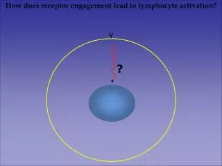
A recognition problem: How do you find the lymphocyte with the correct specificity?
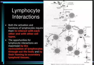
Activation occurs in lymph nodes Lymphocytes are activated in lymph nodes near the site of infection. Following activation, effector T cells migrate to the site of infection, and antibodies are carried through the blood and lymph.
Figure 2-41 Low specificity interactions mediated by chemokines and adhesion molecules direct the trafficking of lymphocytes. Chemokines and chemokine receptors tell lymphocytes where to stop during circulation and where to go within tissues and lymphoid organs.
Cells with the appropriate receptors will move toward the source of a chemokine. Chemokine gradient Movement is mediated by polarized actin reorganization
Adhesion molecules mediate intercellular interactions Intercellular interactions are mediated by adhesion molecules: selectins, integrins, and Ig superfamily proteins
Selectins Selectins recognize specific carbohydrate modifications. Selectin expression is determined by cell type and activation state. Naïve T cells express L-selectin.
Inegrins Integrins are expressed as a and b dimers which can bind a variety of protein ligands, including Ig superfamily members. Integrin activity is controlled by regulated expression and affinity. LFA-1 is one of the integrins expressed on T cells.
Ig Superfamily Ig superfamily members are defined by the presence of a protein domain found in immunoglobulin molecules. Many Ig proteins are ligands for integrins.
T cell homing to lymph nodes or to sites of infection is determined in part by the selective expression and activity of specific chemokines/chemokine receptors and adhesion molecules CCR7 CX3CR3 LFA-1 CXCL10 CCL21
Entry of naïve T cells into the lymph node depends upon three discrete steps: rolling, stopping and diapedesis How does naïve T cell entry into lymph nodes differ from neutrophil recruitment to sites of inflammation?
Boyden chamber/Transwell filter cells chemokine Analysis of C-C chemokine receptor 7(CCR7) -/- mice Forster, R., et al. Cell 99: 23 (1999). (splenocytes) ELC - CCR7 ligand SDF - CXCR4 ligand PBL Spl BM MLN PLN Forster, et al. 1999. Cell 99:23
wild-type wild-type wild-type CCR7-/- Analysis of C-C chemokine receptor 7(CCR7) -/- mice Figure 2 Figure 5 Forster, et al. 1999. Cell 99:23
Within the lymph node, naïve T cell interaction with antigen presenting cells is initiated through interaction of adhesion molecules
TCR signals enhance integrin binding Affinity of integrin interaction is enhanced following successful antigen receptor engagement - “inside-out” signaling
Specificity of T lymphocyte activation relies upon TCR recognition of MHC-peptide: a relatively low affinity interaction compared to immunoglobulin. TCR MHC- peptide MHC I TCR
TCR binds both MHC and peptide The T cell antigen receptor (TCR) recognizes determinants on both the specific peptide and the MHC molecule
CD4/8 coreceptors CD4 and CD8 molecules act as “co-receptors”: they recognize MHC II or MHC I, and help stabilize the TCR-MHC interaction by forming a tripartite complex
CD28 provides 2nd signal Additional receptor-ligand interactions, e.g. CD28 - B7, enhance T cell activation following APC binding.
Molecular rearrangements w/synapse formation T cell receptor engagement with MHC-peptide induces reorganization of cell surface molecules.
Synapse w/scanning EM Re-organization of molecules on the cell surface generates an “immune synapse” which can be analyzed by fluorescence microscopy
Figure 8-29 Immune synapse formation allows the directed release of effector molecules -cytokines and cytotoxic granules, as well as providing a stable interaction with APCs necessary for activation of naïve T cells. Cytoskeleton: green Lytic granules: red
T cell Using fluorescently labeled ligands to assess receptor partitioning following antigen receptor engagement The mature immunological synapse. Patterns of LFA-1 ( A), TCR ( B), and CD28 ( C) interaction in a functional synapse between a T cell and a supported planar bilayer containing Cy5-ICAM-1, Oregon Green-I-A k, and Cy3-B7. All three markers are overlaid in panel D with the cSMAC and pSMAC labeled.SMAC: supramolecular activation complex Bromely, et al. 2001. Ann. Rev. Imm. 19:375
Question for discussion You have identified two new vertebrate organisms with novel immune system anatomy, the first organism has only has a single secondary lymphoid organ, while the second is identical in size to humans but has ten times the number of lymph nodes. A). In the organism with a single secondary lymphoid organ, naïve T cells do not circulate. This organ is identical in structure and function to a human lymph node. Naïve T cells are distributed normally within the organ, and it is the site of lymphocyte activation. However, evolution has led to changes in naïve T cell surface molecules involved in circulation –functions that are no longer required have been lost. What do you expect will have happened to the expression of L-selectin, chemokine receptor CCR7, and integrin LFA-1? For each case explain your answer. B). The second organism, with many more lymph nodes than humans, has an innate and adaptive immune system that uses the same mechanisms and has the same number of cells as in humans. What impact do you expect the increased number of lymph nodes to have on the initiation of an adaptive immune response? Explain your answer.