Download
1 / 22
230 likes | 412 Vues
Molecular Luminescence Spectroscopy. Lecture 31. The relation F = KP 0 e bc implies the following a. The fluorescence signal can be increased if the radiant power of the incident beam is increased. Therefore, always use more intense sources.

E N D
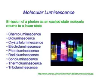
Molecular Luminescence Spectroscopy Lecture 31
The relation F = KP0ebc implies the following a. The fluorescence signal can be increased if the radiant power of the incident beam is increased. Therefore, always use more intense sources. b. The fluorescence signal is directly related to the molar absorptivity and thus molecules of higher molar absorptivities are better fluorescers. c. Fluorescence signal is directly proportional to path length. d. Fluorescence signal is directly proportional to concentration. This is different from relation between absorbance and concentration which is logarithmic. e. The linear correlation between fluorescence and concentration is only true when the absorbance is less than 0.05.
Deviation from Linearity between Fluorescence and Concentration Negative deviations from the linear relation between fluorescence and concentration may be observed in the following cases: a. At absorbances higher than 0.05. b. Self-quenching whereby excited molecules lose their energies by collision with other molecules or solvent c. Self-absorption which occurs when an emission band overlaps with an excitation (absorption) band. In this case, emitted photons excite other molecules in the ground state which results in no net emission.
Fluorescence Instruments Like any emission instrument, a fluorescence or phosphorescence instrument consists of a source, wavelength selectors, a sample cell, a detector, as well as a signal processor. Two wavelength selectors are used, the first is the excitation filter or monochromator which excites the sample while the other is the emission filter or monochromator that separates the fluorescence or phosphorescence wavelength.
Sources We have seen earlier that the fluorescence signal is proportional to the radiant power of the source. Therefore, it is very important to select a source of as high radiant power as possible. In most instruments, a xenon arc lamp is usually the source of choice. However, some instruments use lasers.
Xenon Arc Lamp A quartz envelope hosting two electrodes and xenon at high pressure (5-10 atm). The discharge which takes place between the two electrodes excites xenon and produces a continuum in the range from 200-1000 nm. The wavelength maximum occurs at about 500 nm. The radiant power produced by a xenon arc lamp is very high which makes the lamp suitable for luminescence analysis. Also, the coverage of the whole UV-Vis range adds to the assets of the lamp. However, a very good regulated power supply is essential since any fluctuations of the radiant power will be directly reflected on the fluorescence signal.
Lasers We have seen examples of lasers early in this course. However, it should be indicated that since lasers may have very high intensities, some molecules, which are otherwise non fluorescents, show good fluorescence and can thus be determined by fluorescence spectroscopy. This is called laser induced fluorescence (LIF) and is a common technique in fluorescence spectroscopy. In addition, one should remember that as the radiant power is increased, photodissociation may become a problem especially at shorter excitation wavelengths.
1. Fluorometers When the wavelength selectors are filters, the instrument is called a fluorometer. It is a simple instrument that is usually used for quantitative analysis. The use of fluorometers implies that the excitation and emission wavelengths are predetermined by other means or are known from literature. The detector is usually a sensitive photomultiplier tube. A schematic of a simple fluorometer is shown below:
Unlike the cell used in UV-Vis absorption studies, the cell used in luminescence studies has the two transparent faces perpendicular to each other, rather than parallel. This is important since luminescence is collected 90o to the incident beam. This configuration decreases the noise and fully excludes interferences from the incident beam.
2. Spectrofluorometers In this case, the excitation and emission wavelengths are selected using dispersive elements like gratings or prisms. The same instrumental configuration as fluorometers is usually used. The following schematic represents a basic configuration of a spectrofluorometer:
Excitation and Emission Spectra The first step in a successful fluorescence measurement is to determine the emission wavelengths. The emission spectrum is determined by adjusting the excitation monochromator at an arbitrary, but well selected, wavelength which is shorter than the anticipated emission wavelength. Keeping the emission monochromator at the obtained emission wavelength, the excitation spectrum is recorded by scanning the excitation monochromator in a range less than the emission wavelength of the emission monochromator.
After obtaining the excitation spectrum, the lex (resulting in highest absorbance) is determined. lem is the wavelength with highest fluorescence signal. At this point we are ready to perform a fluorometric determination using the defined excitation and emission wavelengths.One can also start by collecting an absorption spectrum on a UV-Vis instrument. This will give approximate lex to start with, followed by collecting the emission spectrum. Then, after finding lem, one can collect the excitation spectrum to get lex.
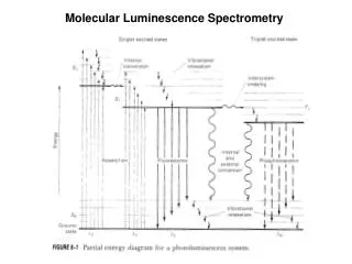
The shape of the emission spectrum is expected to be a mirror image of the excitation spectrum since they originate from opposite processes However, instrumental artifacts result in excitation and emission spectra that are not exactly mirror images.