Download
1 / 42
420 likes | 562 Vues
Reproductive Anatomy. Ex 42. Male Reproductive Anatomy. Penis. Passageway for semen & urine Composed of: bulb body three types of erectile tissues crura attach to pubic arch glans prepuce (foreskin). Penis. Erectile Tissue corpora cavernosum paired tissues laterally

E N D
ReproductiveAnatomy Ex 42
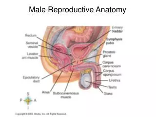
Penis • Passageway for semen & urine • Composed of: • bulb • body • three types of erectile tissues • crura • attach to pubic arch • glans • prepuce (foreskin)
Penis • Erectile Tissue • corpora cavernosum • paired tissues laterally • bound by tunica albuginia • corpora spongiosum • surrounds urethra from bulb to glans
Scrotum • sac of skin and fascia that contains the testis • divided in two by septum • keeps testis outside the abdominal cavity • maintains ideal temperature for spermatogenesis • 3 degrees lower than body temperature
Spermatic Cord • All structures passing to and from the testes • testicular artery • pampiniform plexus of veins • autonomic nerves • lymphatic vessels • ductus (vas) deferens • cremaster muscle
Inguinal Canal & Inguinal Hernias • Inguinal canal is 2 inch long tunnel passing through the • 3 muscles of the anterior abdominal wall -- weakens wall • originates at deep inguinal ring and ends at superficial ring • More common in males
Testicles • Paired organs in scrotum • contains tubes for making and moving sperm and interstitial tissue for making the testosterone • surrounded by fibrous capsule called tunica albuginia • septae divide the tubules into lobules
Testicles • Progression of Tubes • seminiferous tubules • produce sperm • straight tubule (tubulus rectus) • rete testis • efferent ducts (ductule) • epididymis
Testicles • Seminiferous Tubules • spermatogonia and developing sperm • transports sperm to the straight tubule
Testicles • Sertoli (sutentacular) cells • provide environment for developing sperm • blood - testis barrier • sensitive to FSH • secretes • androgen binding protein • concentrates testosterone in tubules • inhibin • released when too many sperm produced • inhibits FSH and GnRH release
Histology • Testicle • sertoli cells • “nurse cells” for sperm • has a prominent nucleolus • spermatogonia • at base of seminiferous tubule
Testicles • Leydig (interstitial cells) • produce testosterone • sensitive to lutienizing hormone • negative feedback with anterior pituitary
Epididymis • Epididymis • attached to posterior part of testis • storage and maturation area for sperm • lined with pseudostratified columnar epithelium with stereocillia • nonmotile stereocillia used to reabsorb fluid and pass nutrients to sperm • surrounded by a thin layer of smooth muscle • moves sperm to ductus deferens (vas deferens)
Ductus Deferens • Tube taking sperm from epididymis to pelvic cavity • thick muscular wall to move sperm • runs over the ureter • forms the ampulla prior to joining the seminal vesicle at the ejaculatory duct
Accessory Glands • Seminal Vesicles • posterior bladder • joins the ductus deferens to form ejaculatory duct • secrets seminal fluid • 60% of volume • fructose • energy for sperm • fibrinogen • clots semen so it can be propelled into vagina • prostaglandins • causes uterine contractions, thins cervical mucous
Accessory Glands • Prostate • gland under bladder • encircles urethra • gland secretions enter in the prostatic urethra • secretions include • citrate • energy source that enters the Krebs (TCA) cycle • proteolytic enzymes • decoagulate the sperm so they can begin to travel
Accessory Glands • Bulbourethral (Cowper’s) Glands • pea-sized glands under the prostate • produces alkaline mucous prior to ejaculation • neutralizes acidic environment in urethra
External Female Genitalia • Mons Pubis • fatty pad • Labia majora/minora • folds of skin • make up vestibule • vestibule • area containing urethra and vagina • clitoris • erectile tissue • bulb of vestibule • erectile tissue • perineum • area between vagina and anus
Vagina • Between rectum and bladder • connection between cervix and exterior • fornix • superior part of vagina surrounding cervix • wall is made up of • adventitia • fibroelastic • muscularis • smooth muscle • mucosa • stratified squamous
Bartholin’s (Vestibular) Glands • located on each side of the vaginal opening • secrete fluid to lubricate vagina • can become obstructed forming cysts (Bartholin’s cysts)
Cervix • inferior “neck” of the uterus • projects into vagina • cervical canal • space between vaginal cavity and uterine cavity • external os • internal os • cervical glands • secrete mucous which blocks sperm entry except at ovulation
Uterus • Muscular organ anterior to rectum and posteror-superior to the bladder • 3 parts • fundus • top part above tubes • body • isthmus • narrowed area between body and cervix • broad ligament • sheets of peritoneum
Uterus • Histology • endometrium • simple columnar epithelium sitting on stroma • stratum basalis • stratum functionalis • built up and shed during menstrual cycle • myometrium • 3 layers of smooth muscle • perimetrium • visceral peritoneum
Uterine (Fallopian) Tubes • Receives ovulated oocyte • Area where fertilization occurs • about 24 hours after ovulation • takes about 7 days for zygote to travel • cilliated simple columnar epithelium and peristalsis • Parts • fimbrae • sweep oocyte into tube • ampulla • body • isthmus
Ovary • Paired organs responsible for • production of oocytes • hormone production • estrogen • progesterone • others • made up of • tunica albuginia • stroma • cortex • site of oogenesis • medulla • ovarian ligament • suspensory ligament
Ovary • Follicles • early oocytes are halted in development • oocyte • centrally located • granulosa cells • hormone producing • if one cell layer thick it is called follicular cells • thecal cells • hormone producing
Ovary • Follicular Development • initiated by FSH • primordial follicle • surrounded by simple squamous follicle cells • primary follicle • 2 or more layers of cuboidal granulosa cells • secondary follicle • fluid filled antrum forms in granulosa cells • graafian follicle • mature follicle, bulges at edge of ovary • corpus luteum • remnants of follicle after ovulation
Ovary • Parts of a follicle • secondary oocyte • zona pellucida • granulosa cells • stalk • corona radiata • antrum • thecal cells
Ovary • Ovulation • surge of LH causes ovulation • release of oocyte from graafian follicle • ovum released into uterine tube • remaining corpus luteum • secrets estrogen and progesteron to maintain the stratum functionalis
Breasts • Modified sweat glands • made up of fat and acini/ducts • fat determines size of the brease • acini produce milk • carried to nipple via lactiferous ducts and lactiferous sinuses • areola • suspensory ligaments • support breast from the deep pectoral facia • lymphatic drainage to axilla
Breasts • Hormonal control • prolactin • stimulates production in presence of estrogen and progesterone • oxytocin • stimulates milk let-down • causes contraction of smooth muscle around acini