Brain Neoplasms: General Considerations
Brain Neoplasms: General Considerations. 1. Comprise: 10% of all tumors 2. Most common childhood neoplasms 3. Peak incidence at 5th decade 4. Supratentorial tumors in adults 5. Infratentorial tumors in childhood. Brain Neoplasms: General Considerations.


Brain Neoplasms: General Considerations
E N D
Presentation Transcript
Brain Neoplasms:General Considerations 1. Comprise: 10% of all tumors 2. Most common childhood neoplasms 3. Peak incidence at 5th decade 4. Supratentorial tumors in adults 5. Infratentorial tumors in childhood
Brain Neoplasms:General Considerations 6. Different tumors in different ages 7. Primary tumors infiltrative, metastatic well-demarcated 8. Intraneural seeding occur, but no extraneural metastasis 9. Produce neurologic symptoms by size, location, invasiveness, and secondary effects
Varieties of brain tumors • Meninges: meningioma, hemangiopericytoma • Astrocytes: astrocytoma (various types) • Oligodendrocytes: oligodendroglioma • Ventricles: ependymoma, choroid plexus papilloma, colloid cyst • Vascular: hemangioblastoma • Primitive cells: germinoma, medulloblastoma, neuroblastoma, pineoblastoma, retinoblastoma • Neuronal: ganglioglioma, gangliocytoma • Pituitary: adenoma, craniopharyngioma • Nerves: schwannoma, neuroblastoma
Incidence of Intracranial Gliomas(All ages) Glioblastomas Astrocytomas Ependymomas Medulloblastomas Oligodendrogliomas Choroid plexus papillomas Colloid cysts 55.0% 20.5% 6.0% 6.0% 5.0% 2.0% 2.0%
Incidence of Primary IntraspinalIntramedullary Gliomas 63.0% 24.5% 7.5% 3.0% 2.0% Ependymomas Astrocytomas (grades 1 and 2) Glioblastomas (Astrocytomas grades 3 and 4) Oligodendrogliomas Other tumors
Frequent brain tumors • Meningioma • Astrocytoma/glioblastoma • Oligodendroglioma • Ependymoma • Medulloblastoma • Schwannoma/neurofibroma • Phakomatosis
Meningioma Arachnoid cells origin Attached to dura, subdural Common sites Changes in cranium Hyperostosis Invasion Microscopic: whorls and psammoma bodies
Gliomas • Astrocytes- astrocytomas • Fibrillary • Pilocytic • Oligodendrocytes- oligodendrogliomas • Ependyma- ependymomas
Astrocytomas Adults: Childhood: Supratentorial Solid Malignant Infratentorial Cystic Benign
Adult vs childhood astrocytomas • Adult: fibrillary. Grading varies from low grade malignancy to one of most malignant brain tumor. • Childhood: pilocytic. Very low grade tumor (benign).
Fibrillary astrocytomas • Grossly solid • Common in cerebral hemispheres • Low grade in young, higher grade in older • Grading • astrocytoma (low grade) • Anaplastic astocytoma • glioblastoma multiforme
Fibrillary astrocytoma: microscopic • Low grade- hypercellularity, pleomorphism • Anaplastic- as above plus mitosis, vascular endothelial proliferation • Glioblastoma multiforme- as above plus necrosis and pseudopalisades. Grossly variegated appearance (multiforme)
Pilocytic astrocytoma • Common in childhood • Most slow growing of the gliomas • Sites: cerebellum, around III V., optic nerve • Grossly cystic with mural nodule • Microscopic • elongated hair-like (pilo) elongated cells • Rosenthal fibers
Rosenthal fiber definition • Dense, eosinophilic fibers within cytoplasmic processes of astrocytes. • Correspond to aggregate accumulation of intermediate filaments in these processes.
Oligodendroglioma Slow growing tumor Potentially malignant Calcifications
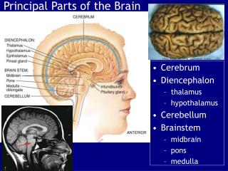
Tumors in Ventricles 1. Ependyma: Ependymoma 2. Choroid Plexus: Papilloma
Ependymomas • Arise from ependymal lining- ventricles and central canal of spinal cord • Common in childhood • 4th V. common in cerebrum • Most common tumor of spinal cord parenchyma in adult • Microscopic • perivascular pseudorosettes • ependymal rosettes
Primitive neuroectodermal tumors • Neuroblastoma- cerebral hemispheres • Medulloblastoma- cerebellum • Ependymoblastoma- ventricles • Pineoblastoma- pineal region
Medulloblastoma • Origin: primitive neuroectodermal cells • Age: 1st decade of life • Site: vermis of cerebellum • May cause hydrocephalus • Subarachnoid dissemination
Histologic patterns: definitions • Whorls: onion-skinning pattern of tumor cells • Psammoma bodies: laminated calcium • Pseudopalisading: lining up of the tumor cells around a central necrotic area • Palisade: lining up of tumor cells around their own cytoplasmic processes. No necrosis. • Pseudorosette: tumor cells around blood vessels, cells equidistant from vessel walls. • Rosettes: tumor cells around central lumen or fibrillary area of cellular processes
Brain Tumors: Microscopic Meningioma Whorls and psammoma bodies Glioblastoma Pseudopalisades Oligodendroglioma Mosaic/poached-egg Ependymoma Perivascular pseudorosettes Medulloblastoma Rosettes
Tumors of Nerve Rootsand Peripheral Nerves 1. Schwannoma viii Cranial nerve (Acoustic sch.) Spinal roots, posterior Peripheral nerves 2. Neurofibroma Spinal Roots, rare Peripheral nerves 3. Malignant variants Rare
Peripheral nerve tumors Neurofibroma • Schwann cells, neurites, fibroblasts • Fusiform and involves nerve trunk • Not encapsulated • Not resectable without sacrificing nerve • Micro- Intermingled cells with wavy nuclei Schwannoma • Schwann cells • Compress the nerve trunk • Encapsulated • Easily resectable without nerve damage • Microscopic: • Antony A and B fibers • Verocay bodies
Metastatic brain tumors • Most common brain tumor in adults. • Common primary sites: melanoma, lung, breast, GI tract, kidney. • Most are in cerebrum (MCA territory). • In gray-white junctions due to rich capillarity • Discrete, globoid, sharply demarcated tumors. Amenable to surgical resection. • Single or multiple. • Brain edema frequent.
Phakomatosis: definition • Phakos (Greek): lentil mole or freckle. • Neurologic abnormalities combined with defects of skin or retina, explained by their common ectodermal origin. • Involvement of visceral organs
Phakomatosis(Neurocutaneous dysplasia) 1. Neurofibromatosis (von Recklinghausen's dis.) 2. Tuberous Sclerosis 3. Sturge-Weber disease (Encephalofacial Angiomatosis) 4. von Hippel-Lindau Disease 5. Neurocutaneous Melanosis
Neurofibromatosis 1. Dominant inheritance 2. Multiple neurofibromas Central - CNS peripheral nerves 3. Increased incidence of: meningioma glioma schwannoma - bilateral VIII N. 4. Cafe-au-lait (melanosis) in skin 5. Elephantiasis: increased connective tissue
Tuberous Sclerosis 1. Dominant inheritance 2. Clinical triad: seizures mental retardation adenoma sebaceum 3. Retinal hamartoma (phakoma) 4. Tubers in cerebral cortex 5. Subependymal giant cell astrocytoma 6. Hamartomas in other organs: heart, kidney