Understanding RNA Types and Their Secondary Structures in Bioinformatics
290 likes | 432 Vues
This overview delves into the diverse types of RNA, including coding mRNA, non-coding rRNA, tRNA, snRNA, siRNA, and ribozymes. It explores methodologies for identifying RNA genes and emphasizes the importance of secondary structure, such as in transcription regulation and RNA interference. The analysis methodologies, such as dynamic programming for RNA secondary structure prediction using tools like Mfold, are discussed. Insights into the role of siRNAs in gene silencing and their implications for gene targeting are also highlighted, showcasing their significance in modern bioinformatics research.

Understanding RNA Types and Their Secondary Structures in Bioinformatics
E N D
Presentation Transcript
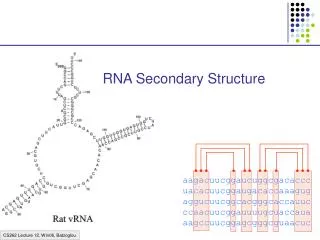
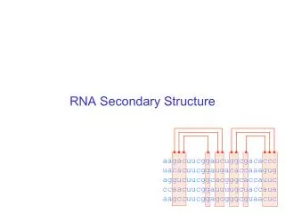
RNA BioinformaticsGenes and Secondary Structure Anne Haake Rhys Price Jones & Tex Thompson
Types of RNA • Coding • mRNA (messenger) • Non-Coding • rRNA (ribosomal) • tRNA (transfer) • snRNA (small nuclear;splicing) • RNAseP (ribozyme) • siRNA (small inhibitory) • Others…
RNA genes? Recall: • Protein-coding genes; we have relatively good methods • Ab initio • Homology-based • RNA genes • Poor sequence homology • Secondary structure useful
Functional Roles: RNA Secondary Structure • mRNA • Regulation of transcription termination • Regulation of translation initiation • rRNA • ribosomal structure • tRNA • adaptor in translation • RNA interference • Regulation of gene expression • Anti-viral activity
mRNAs have functional secondary structures • e.g. Transcription Termination Signal
Ribozymes: Enzymes made of RNA • RNA molecules in Tetrahymena were shown to splice out introns without the aid of proteins • Ribozymes have been discovered in higher organisms, and may play a role in processing mRNA
Ribonuclease P • Enzyme found in many organisms, cleaves the 5’ end of tRNA molecules • Heterodimer consisting of a protein molecule and an RNA molecule • Without RNA molecule, Ribonuclease P loses all activity • Without protein, Ribonuclease P shows only reduced activity
Pictures from the Web • http://www.mbio.ncsu.edu/RNaseP/rna/threed/threed.html • http://proteinexplorer.org (molecule 1d6t)
Discovery of Ribonuclease P http://www.mun.ca/biochem/courses/3107/Lectures/Topics/Splicing.html
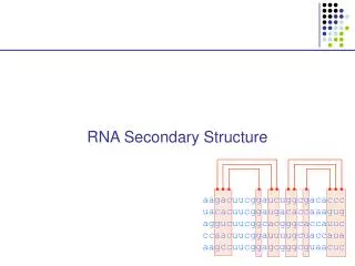
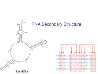
Can identify RNA genes that belong to a known family • Infer secondary structure by comparing sequences (multiple alignments) • e.g. Look for covariance; positions that covary to maintain Watson-Crick base-pairing;implies role in secondary structure • Rfam: a collection of multiple alignments and covariance models for ncRNAs • Rfam
Prediction of RNA Secondary Structure • Find the configuration that maximizes the number of base pairs • Scoring all possibilities would be computationally expensive • Use dynamic programming • Thermodynamics approach • Mfold: uses an energy minimization method of Zuker • http://www.bioinfo.rpi.edu/applications/mfold/ • http://bioweb.pasteur.fr/seqanal/interfaces/mfold-simple.html
RNA Interference • Breakthrough of the year in 2002 • Discovered in C. elegans • dsRNA involved in sequence-specific gene silencing • Post-transcriptional gene silencing • 21-25 nucleotide dsRNAs (siRNAs) facilitate the degradation of homologous RNAs
RNAi • Useful for gene targeting to study function • Other techniques for gene targeting • “knock-out” by homologous recombination • Antisense • siRNA-direct “knock-down” has potential to allow systematic study of each gene in a pathway • siRNA might allow silencing of pathogenic genes or pathogens (e.g. viruses)
Mechanism • siRNAs: 21-23 nt dsRNA with 2-3 nt 3’ overhangs • Produced from cleavage of long dsRNAs by “Dicer” enzyme • Form a siRNA-protein complex “RISC” • Cleaves homologous mRNA target • Also can start with a hairpin precursor rather than dsRNA
siRNA demo • RNA Interference links and refs
Introduction to Proteomics Techniques & Computational Issues
Experimental Techniques • As with transcriptome analysis, proteome analysis is limited by the techniques currently available • But, proteome analysis even more difficult and less precise due to the nature of proteins
Two-dimensional Gel Electrophoresis • 2D gels • First dimension: isoelectric focusing • Separates proteins on basis of charge • Second dimension: SDS-PAGE • Able to resolve thousands of proteins in a single gel • Proteins are usually radioactively labeled
Challenges of 2D gel Analysis • Reproducibility • Software is available to assist in aligning the spots between gels and integrating the intensities of the spots • Identification of the proteins of interest • Some underrepresented e.g. membrane proteins • Some below levels of detection • Which protein is represented by each spot? • Mass spectrometry has greatly enhanced ability to identify individual proteins
2-Dimensional Gel Electrophoresis • http://us.expasy.org/ • For other examples and tools
Mass Spectrometry • Able to uniquely identify the proteins associated with individual spots in 2D gels • Spots are excised from gels • Proteins are digested into peptide fragments using proteases such as trypsin • Trypsin cleaves peptide bonds next to the amino acids lysine and arginine. • Peptides are ionized for Mass Spec analysis • For a quick explanation of Mass Spec see: http://www.jeol.com/ms/docs/whatisms.html
Mass Spectrometry • Generates a peptide mass fingerprint • Computational challenge: the fingerprint must be matched up with the theoretical mass spectrum of the proteins derived from genomics databases • Analysis software ProteinProspector
Protein Microarrays • High-throughput techniques similar to gene chips • Probes (e.g. antibodies) attached to chips • Fluorescently-labeled proteins washed over chips • Fluorescence intensity indicative of relative levels • Variations include protein-compound (drug) interactions, protein-DNA etc.
Protein Microarrays • Major problems with analysis of proteins in this way • Protein-protein binding not determined by strict rules as it is in nucleic acids (base-pairing) • One protein may bind several others on the chip • Protein interactions very sensitive to chemistry • Application of protein arrays often used as a follow-up to gene chip studies