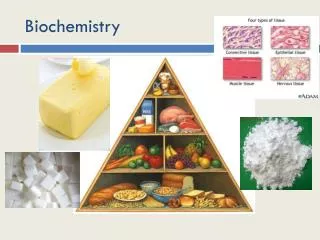
Biochemistry
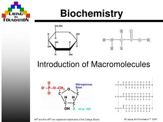
Biochemistry. Robert F. Waters PhD. Porphyrin Rings. Produced mainly in: Liver Erythrocyte producing cells of bone marrow Not mature erythrocytes (lack of mitochondria) Initial step and last three steps are in the mitochondria Glycine and Succinyl CoA are precursors

Biochemistry
E N D
Presentation Transcript
Biochemistry • Robert F. Waters PhD.
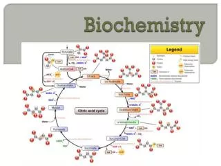
Porphyrin Rings • Produced mainly in: • Liver • Erythrocyte producing cells of bone marrow • Not mature erythrocytes (lack of mitochondria) • Initial step and last three steps are in the mitochondria • Glycine and Succinyl CoA are precursors • Hemin is feedback inhibitor of -aminolevulinic dehydrase (ALA) (-delta) • Lead is inhibitor of this pathway
Porphyrin Uses • Cytochromes • P450, b5, ETS • Hemoglobin
Porphyrin Pathway • Begins in Mitochondria • Vitamin necessary in first enzyme reaction • Pyridoxal Phosphate • Porphobilinogen is first molecule in cytosol
Porphyrins Cont: • Addition of ferrous iron in mitochondria • ferrochelatase
Porphyrias • Usually hereditary porphyrin production defects • General Classification • Erythropoietic (defect in RBCs) • Hepatic (Defect in liver) • Usually autosomal dominant • Covered in genetics
Heme Degradation • First product is biliverdin (open ring) • Heme oxygenase • Biliverdin plus CO yields bilirubin • Biliverdin reductase • NADP • Bilirubin • Gut (microbial enzymes act on bilirubin) • Produce Urobilinogen • Absorbed and carried to liver to produce Urobilin • Yellow color • Large intestine further microbial enzymes produce Stercobilin • Characteristic brown color • Liver • Conjugation with 2 moles of glucuronic acid • Glucuronyl bilirubin transferase • Forms bilirubin diglucuronide (polar and soluble) • detox
Jaundice • Hemolytic • Response to sickle cell anemia • Glycolytic enzyme deficiencies • Erythroblastosis foetalis • Obstructive • Hepatic tumor • Pale stools • GI pain, nausea • Hepatocellular • Liver damage • Cirrhosis, hepatitis • Urine dark, stools pale (liver regurgitates conjugated bilirubin into blood and then into urine) • Elevated AST (SGOT) and ALT (SGPT)
Hemoglobin • Porphyrin ring • Iron • Oxygen Binding • Two alpha chains and two beta chains • 1212 • Thalassemias
Genetics of Hemoglobin • 12 • Chromosome 16 • Diploid designation 12/12 • Produced in utero • 12 • Chromosome 11 • Produced postpartum only • Alpha-like chains () • pre- & post-natal • (zeta) • Beta-like chains () • Essentially post-natal • (sigma, epsilon, gamma)
Hemoglobin Oxygen Release • High acidity causes hemoglobin to release oxygen • Erythrocytes passing through tissue that are producing acids-lactic acid • Handoff to myoglobin • Called Bohr effect (Christian Bohr-Physiologist) • Named after father of noted physicist Niels Bohr • 2,3-bisphosphoglycerate promotes release of oxygen by hemoglobin
Cooperative Oxygen Binding • Myoglobin-rectangular hyperbola • Hemoglobin-sigmoidal • Partial pressure • Saturation
Carbon Dioxide & Hb • Isohydric Transport of CO2 • Gas exchange without pH change • Carbonic anhydrase (Zn-containing)
Another Mechanism of CO2 Transport • Direct reaction of carbon dioxide to produce carbaminohemoglobin
Genetics Overview • -psi pseudo-genes • Mutations (mutated) such that they do not produce a functional protein • -zeta
Hemoglobinopathies • Very common • AR-Sickle Cell Anemia • HbA vs HbS (6 GluVal) • Life long hemolytic anemia
Hemoglobinopathies-Cont: • Thalassemias (thalassa-sea:many cases around Mediterranian Sea) • + 0 (some production vs. none) • + 0(some production vs. none) • Alpha thalassemias affect fetal and postpartum hemoglobin • Beta thalassemias affect only postpartum
Thalassemias-Cont: • Alpha thalassemia usually more severe • Thalassemia major • Variety of deletions (usually) • Beta thalassemia usually less severe • Thalassemia minor • Usually single nucleotide substitutions
Iron • Association with copper • Absorption from lumen in intestine • Ceruloplasmin • Cupric to cuprous, ferrous to ferric • Vitamin C • Wilson’s disease • 1:100,000 • Lack of copper transport proteins
Iron Contained in:Some Examples • Hemoglobin, myoglobin • NO binding, guanylate cyclase • ETS hemes • Cytochrome b5 in desaturation • Iron-sulfur (Complex I, aconitase, xanthine oxidase, ferrochelatase (heme synth.) • Phenylalaine hydroxylase, tyrosine hydroxylase, dioxygenases • Etc.
Proteins and Iron • Iron binding proteins • Transferrin (Fe3+), Lactoferrin (Fe3+), Ferritin (Fe3+), Hemosiderin (Fe3+) • Proteins that use iron as substrate • Ferroxidase (Fe2+/Fe3+)-adrenals, Ferrochelatase (Fe2+)-porphyrins • Protein that uses heme as substrate • Heme oxygenase (biliverdin)
Transferrin • Plasma protein • Glycoprotein synthesized by liver • Single polypeptide (~700 AA’s) • High affinity for ferric iron • No affinity for ferrous iron • Serum levels about 30umol/L • Serum has excess iron binding capacity
Transferrin Cont: • Transferrin production increased during • Iron deficiency • Pregnancy (high estrogen levels) • Women taking oral contraceptives • Transferrin production decreased • Excess iron • Infection • Inflammation • Neoplasia • Protein catabolic state • Transferred to recipient cells by- • Transferrin-binding Receptors
Ferritin • Store iron in ferrous non-toxic state • Relatively short term storage • Handoff to/from transferrin • Handoff to/from hemosiderin • Mainly intracellular • Not usually in the serum unless iron storage saturation
Other Transport Proteins • Exist during high iron overload, ineffective erythropoiesis (hemes/hemoglobin in serum), hemolytic anemia, etc. • Examples • Haptoglobin • Bind free (serum) oxyhemoglobin dimers • Brought into hepatocytes by receptor mediated endocytosis • Hemopexin and albumen bind free hemes • Lactoferrin (neutrophils, secretory epithelial secretions (milk)
Hemosiderin • Long term storage • Handoff to/from ferritin • Exist in times of iron overload • Probably a form of iron-ferritin complexes in a type of micelle formation in tissues
Hypochromic Anemia • Pale RBCs due to low levels of hemoglobin
Hemochromatosis • Excess iron (free iron due to saturation of tranferrin) • Arthritis, liver cancer, coronary occlusions • Early diagnosis and treatment • AR inheritance with gene on chromosome 6 • Treatment • Venesection (removal of blood) • Removal of 500ml of blood over specified frequency and period of time to lower iron reserves • High frequency is 500ml per week over 1-2 year period of time • Chelation
Assessment of Iron • Serum Ferritin Concentration • Quantitative relationship to iron stores • 20-200ng/mL • Remember:ferritin is usually tissue bound • Note: Some research has shown a relationship between excess iron, and heart disease and cancer • Supplement only when deficient