Download

1 / 26
260 likes | 512 Vues
The pharynx Dr.Nimir Dr.Safaa. Objectives. Define Pharynx. Describe its different parts. Why its anterior wall is deficient. Discuss the pharyngeal muscles. Give its blood supply and venous drainage. Discuss its sensory and motor nerve supply. The pharynx(throat):

E N D
The pharynx Dr.Nimir Dr.Safaa
Objectives • Define Pharynx. • Describe its different parts. • Why its anterior wall is deficient. • Discuss the pharyngeal muscles. • Give its blood supply and venous drainage. • Discuss its sensory and motor nerve supply.
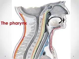
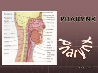
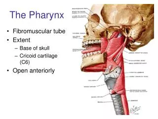
The pharynx(throat): The pharynx is attached above to the base of the skull and is continuous below, approximately at the level of vertebra CVI, with the top of the esophagus. The walls of the pharynx are attached anteriorly to the margins of the nasal cavities, oral cavity, and larynx. It is subdivided into three regions, the nasopharynx, oropharynx, and laryngopharynx. The posterior apertures (choanae) of the nasal cavities open into the nasopharynx. The posterior opening of the oral cavity (oropharyngeal isthmus) opens into the oropharynx.
The superior aperture of the larynx (laryngeal inlet) opens into the laryngopharynx. The pharyngotympanic tubes open into the lateral walls of the nasopharynx. Lingual, pharyngeal, and palatine tonsils are on the deep surface of the pharyngeal walls. The pharynx is separated from the vertebral column behind by a thin retropharyngeal space containing loose connective tissue.
The muscles of the pharynx are organized into two groups: 1.Constrictor muscles: The three constrictor muscles on each side are: Superior, middle, and inferior constrictor muscles. Posteriorly, the muscles from each side are joined together by the pharyngeal raphe. Anteriorly, these muscles attach to bones and ligaments related to the lateral margins of the nasal and oral cavities and the larynx. The constrictor muscles overlap each other. Collectively, the constrictors constrict or narrow the pharyngeal cavity. All of the constrictors are innervated by the pharyngeal branch of the vagus nerve [X].
2.The three longitudinal muscles of the pharyngeal wall are: Stylopharyngeusfrom the styloid process, salpingopharyngeus from the cartilaginous part of the pharyngotympanic tube, and palatopharyngeus from the soft palate. From their sites of origin, these muscles descend and attach into the pharyngeal wall. The longitudinal muscles elevate the pharyngeal wall. Salpingopharyngeusand palatopharyngeus are innervated by the vagus, stylopharyngeus by the glossopharyngeal nerve [IX].
Nasopharynx : The nasopharynx is behind the posterior apertures (choanae) of the nasal cavities and above the level of the soft palate. The cavity of the nasopharynx is continuous below with the cavity of the oropharynx at the pharyngeal isthmus. Elevation of the soft palate and constriction of the palatopharyngeal sphincter close the pharyngeal isthmus during swallowing. There is a large collection of lymphoid tissue (the pharyngeal tonsil) in the mucosa covering the roof of the nasopharynx. Enlargement of this tonsil, known as adenoids, can occlude the nasopharynx so that breathing is only possible through the oral cavity.
The most prominent features on each lateral wall of the nasopharynx are: The opening of the pharyngotympanic tube. Mucosal elevations and folds covering the end of the pharyngotympanic tube and the adjacent muscles. Mucosal folds related to the pharyngotympanic tube include: The small vertical salpingopharyngeal fold, which descends from the tubal elevation and overlies salpingopharyngeus muscle. A broad fold or elevation (torus levatorius) overlies the levatorvelipalatini muscle.
Oropharynx: The oropharynx is posterior to the oral cavity. The palatoglossal folds (arches), one on each side, that cover the palatoglossal muscles, mark the boundary between the oral cavity and the oropharynx. The arched opening between the two folds is the oropharyngeal isthmus. Just posterior and medial to these folds are another pair of folds (arches), the palatopharyngeal folds, one on each side, that overlie the palatopharyngeus muscles. The palatine tonsils are on the lateral walls of the oropharynx. They are collections of lymphoid tissue between the palatoglossal and palatopharyngeal arches.
Laryngopharynx : The laryngopharynx extends from the superior margin of the epiglottis to the top of the esophagus at the level of vertebra CVI. The cavity of the laryngopharynx is related anteriorly to a pair of mucosal pouches (valleculae) between the base of the tongue and epiglottis. There is another pair of mucosal recesses (piriform fossae) between the central part of the larynx and the more lateral lamina of the thyroid cartilage.
Arteries that supply upper parts of the pharynx include: The ascending pharyngeal artery. The ascending palatine and tonsillar branches of the facial artery. Arteries that supply the lower parts of the pharynx include pharyngeal branches from the inferior thyroid artery. The major blood supply to the palatine tonsil is from the tonsillar branch of the facial artery, which penetrates the superior constrictor muscle.
Veins of the pharynx form a plexus, which drains superiorly into the pterygoid plexus in the infratemporalfossa, and inferiorly into the facial and internal jugular veins. Lymphatic vessels from the pharynx drain into the deep cervical nodes and include retropharyngeal, paratracheal, and infrahyoid nodes. The palatine tonsils drain through the pharyngeal wall into the jugulodigastric.
Motor and most sensory innervation (except for the nasal region) of the pharynx is mainly through branches of the vagus [X] and glossopharyngeal [IX] nerves. The pharyngeal plexus is formed by: The pharyngeal branch of the vagus nerve [X]. Branches from the external laryngeal nerve from the superior laryngeal branch of the vagus nerve [X]. Pharyngeal branches of the glossopharyngeal nerve [IX]. All muscles of the pharynx are innervated by the vagus nerve [X] mainly through the pharyngeal plexus, except for the stylopharyngeus, which is innervated directly by a branch of the glossopharyngeal nerve [IX].
Each subdivision of the pharynx has a different sensory innervation: The nasopharynx is innervated by a pharyngeal branch of the maxillary nerve [V2] that originates in the pterygopalatinefossa and passes through the palatovaginal canal in the sphenoid bone to reach the roof of the pharynx. The oropharynx is innervated by the glossopharyngeal nerve [IX] via the pharyngeal plexus. The laryngopharynx is innervated by the vagus nerve [X] via the pharyngeal plexus.