
Posterior Abdominal Wall
E N D
Presentation Transcript
Posterior Abdominal Wall Abdomen, Pelvis & Perineum Unit Lecture 3 د. حيدر جليل الأعسم
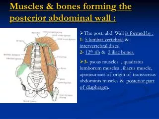
Posterior Abdominal Wall • Bony structures: Five lumbar vertebrae and their intervertebral discs in the midline and the 12th ribs and upper part of the bony pelvis (ilium) laterally B. Muscular structures: psoas muscles, quadratuslumborum muscles, aponeuroses origin of transversusabdominis muscles, posterior part of diaphragm and iliacus muscles that lie in the upper part of the bony pelvis.
Psoas Major Origin:roots of transverse processes, sides of vertebral bodies, and intervertebral discs, from the 12th thoracic to the 5th lumbar vertebrae. Insertion: lesser trochanter of femur. Muscle fibers run downward and laterally and leave the abdomen to enter the thigh behind the inguinal ligament. The psoas is enclosed in a fibrous sheath that is derived from the lumbar fascia. The sheath is thickened above to form the medial arcuate ligament. Nerve supply:by lumbar plexus. Action:flexes the thigh at the hip joint on the trunk, or if the thigh is fixed, it flexes the trunk on the thigh.
Quadratus Lumborum It is a flat, quadrilateral-shaped muscle that lies alongside vertebral column. Origin:iliolumbar ligament, adjoining part of iliac crest, and tips of transverse processes of lower lumbar vertebrae. Insertion:into the lower border of the 12th rib and the transverse processes of the upper four lumbar vertebrae. The muscle fibers run upward and medially and its anterior surface is covered by lumbar fascia, which is thickened above to form the lateral arcuate ligament and below to form the iliolumbar ligament. Nerve supply: This muscle is supplied by the lumbar plexus. Action:It fixes or depresses 12th rib during respiration and laterally flexes vertebral column to the same side.
Iliacus Fan shaped muscle Origin:upper part of iliac fossa. Insertion:lesser trochanter of femur because its fibers join lateral side of psoas tendon. combined muscles are often referred to as the iliopsoas. Nerve supply:femoral nerve, a branch of the lumbar plexus. Action:iliopsoasflexes the thigh on the trunk at the hip joint, or if the thigh is fixed, it flexes the trunk on the thigh.
Fascial Lining of the Abdominal Walls Abdominal walls are lined by one continuous layer of connective tissue that lies between parietal peritoneum and muscles. Fascia is named according to structure that covers it: Diaphragmatic fasciacovering under-surface of diaphragm Transversalis fasciacovering transversus abdominis. Psoas fasciacovering psoas muscle. Quadratus lumborum fasciacovering quadratus lumborum. Fascia iliacacovering iliacus muscle. The blood and lymph vessels lie within this fascial lining, whereas the principal nerves lie outside the fascia.
Femoral sheath Femoral sheathis a downward prolongation of the fascial lining around the femoral vessels and lymphatics, for about 1.5 in. (4 cm) into the thigh, behind the inguinal ligament. Because the femoral nerve lies outside the fascial envelope, it has no sheath.
Surface Anatomy: Surface Landmarks of Abdominal Wall Abdomen, Pelvis & Perineum Unit Lecture 3 د. حيدر جليل الأعسم
Surface Abdominal Landmarks Xiphoid Process: palpated in the depression where costal margins meet in the upper part of the anterior abdominal wall. The xiphisternal junction lies opposite the body of the ninth thoracic vertebra (T9). Costal Margin: curved lower margin of thoracic wall and is formed in front by cartilages of 7th, 8th, 9th & 10th ribs and behind by cartilages of 11th & 12th ribs. The costal margin reaches its lowest level at the 10th costal cartilage, which lies opposite the body of third lumbar vertebra (L3).
Surface Abdominal Landmarks Iliac Crest can be felt along its entire length from anterior superior iliac spine to posterior superior iliac spine. Its highest point lies opposite body of fourth lumbar vertebra (L4) Pubic Tubercle a small protuberance along superior surface of pubis. Symphysis Pubis midline cartilaginous joint Between bodies of the pubic bones and felt as a solid structure beneath the skin in the midline at the lower extremity of the anterior abdominal wall.
Surface Abdominal Landmarks Inguinal Ligament It lies beneath the groin skin crease. It is attached laterally to the anterior superior iliac spine and curves downward and medially, to be attached to the pubic tubercle. Superficial Inguinal Ring The superficial inguinal ring is a triangular aperture in the aponeurosis of the external oblique muscle and is situated above and medial to the pubic tubercle.
Surface Abdominal Landmarks Linea Alba: is a vertically running midline fibrous band that extends from symphysis pubis to xiphoid process. It is formed by fusion of aponeuroses of anterior abdominal wall muscles. Umbilicus: is a puckered scar in linea alba that represent site of attachment of umbilical cord in the fetus. It is inconstant in position. Tendinous Intersections of the Rectus Abdominis: are three in number running across rectus abdominis muscle at the level of tip of xiphoid process, umbilicus and halfway between them. Linea Semilunaris: is the lateral edge of rectus abdominis muscle & crosses costal margin at tip of ninth costal cartilage.
Abdominal Lines and Planes Vertical Lines: Midline, mid-clavicular line Horizontal Planes: Transpyloric Plane : passes through tips of ninth costal cartilages on both sides. It lies at level of body of first lumbar vertebra (L1), pylorus of the stomach, duodenojejunal junction, neck of pancreas, and hila of kidneys. Subcostal Plane : joins lowest point of costal margin on both sides that is 10th costal cartilage & at level of third lumbar vertebra (L3). Intercristal Plane : passes across highest points on iliac crests & lies on level of body of fourth Lumbar vertebra (L4). This is commonly used as a surface landmark when performing a lumbar spinal tap. Intertubercular Plane : joins tubercles on iliac crests & lies at level of fifth lumbar vertebra (L5).
Abdominal Surface Regions • 4 quadrants: use one midline vertical line & one horizontal line that intersect at the umbilicus. Quadrants are upper right, upper left, lower right, and lower left. • 9 regions: use two vertical lines (right & left midclavicular lines) & two horizontal lines (subcostal & intertubercular planes). The regions are: three central regions (epigastric, umbilical & suprapubic) and three lateral regions on each side (hypochondriac, lumber or flank and iliac or groin).
Abdominal Hernias Hernia: is a protrusion of part of abdominal contents beyond normal confines of abdominal wall. It consists of 3parts: sac, contents of the sac & coverings of the sac. A. Hernialsac is a pouch (diverticulum) of peritoneum and has a neck and a body. B. Hernialcontents may consist of any structure found within the abdominal cavity and may vary from a small piece of omentum to a large viscus such as the kidney. C. Hernial coverings: layers of abdominal wall through which hernial sac passes. Abdominal hernia types: • Inguinal (indirect or direct) Hernia • Femoral Hernia • Umbilical (congenital or acquired) Hernia • Epigastric Hernia • Separation of the recti abdominis Hernia • Incisional Hernia • Hernia of lineasemilunaris (Spigelian hernia) • Lumbar (Petit's triangle hernia) • Internal Hernia
Abdominal Hernias Indirect Inguinal Hernia • most common form of hernia • Congenital in origin, hernialsac is the remains of the processusvaginalis. • Hernia sac enter deep inguinal ring and may extend to extends through superficial inguinal ring & down into the scrotum or labium majus. • Neck of the hernial sac lies at deep inguinal ring lateral to inferior epigastricvessels, Body of the sac resides in the inguinal canal and scrotum (or base of labium majus).
Abdominal Hernias Direct Inguinal Hernia • about 15% of all inguinal hernias, rare in women and most are bilateral • Acquired type, It is a disease of old men with weak abdominal muscles. • Hernial sac of bulges directly anteriorly through posterior wall of the inguinal canal medial to inferior epigastricvessels. • Neck of the hernial sac is wide because of presence of strong conjoint tendon, this hernia is usually nothing more than a generalized bulge.
Abdominal Hernias Femoral Hernia Femoral hernia is more common in women. Hernial sac descends through femoral canal within the femoral sheath, creating a femoral hernia. Femoral canal is a compartment for the lymphatics and occupies the medial part of the sheath. Its upper opening is referred to as the femoral ring. Femoral septum is a condensation of extraperitoneal tissue that plugs the opening of the femoral ring. On escaping through the lower end of femoral canal, it expands to form a swelling in the upper part of the thigh. Neck of hernia lies below & lateral to the pubic tubercle at femoral ring; which serves to distinguish it from an inguinal hernia.