Download
1 / 13
140 likes | 294 Vues
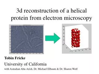
3 d reconstruction of a helical protein from electron microscopy. Tobin Fricke University of California with Asmahan Abu-Arish, Dr. Michael Elbaum & Dr. Sharon Wolf. Agrobacterium tumefaciens. Infection by gene transfer from bacterium to plant. Growth factor Enzymes to produce opines.

E N D
3d reconstruction of a helical protein from electron microscopy Tobin Fricke University of California with Asmahan Abu-Arish, Dr. Michael Elbaum & Dr. Sharon Wolf
Agrobacterium tumefaciens Infection by gene transfer from bacterium to plant Growth factor Enzymes to produce opines Can replace with other genes for transformation Evidence for gene transfer to animal cells
electron microscopy • Just like optical microscopy, but with electrons instead of photons
tomography • by looking through an object at many angles, determine its internal structure.
Helical symmetry • Translation is equivalent to axial rotation
Results: T-complex structure hollow helical tube diameter 16 nm pitch 5.2 nm 4.3 VirE2/turn shortening factor ~7 3-domain structure of VirE2 single-strand DNA lies on a shelf at the interior of the tube at radius ~5.5 nm 20 kb ssDNA: ext. length 8. mm random coil ~90 nm