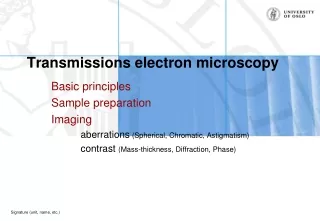
Fundamentals of Electron Microscopy: Lens Aberrations and Corrections
Understand lens aberrations, their significance, and correction methods for optimal electron microscopy resolution. Learn about spherical aberration, chromatic aberration, astigmatism, and TEM column components. Explore electron-matter interactions and imaging challenges.


Fundamentals of Electron Microscopy: Lens Aberrations and Corrections
E N D
Presentation Transcript
Basic Electron Microscopy Arthur Rowe The Knowledge Base at a Simple Level
Introduction • These 3 presentations cover the fundamental theory of electron microscopy • In presentation #2 we cover: • lens aberrations and their importance • how we correct for lens astigmatism • limits to ultimate resolution of the TEM • Interactions of electrons with matter
aberrations of electromagnetic lenses the most important ones to consider are: • spherical aberration • chromatic aberration • astigmatism
spherical aberration object plane • arises because a simple lens is more powerful at the edge than at the centre • is not a problem with glass lenses (can be ground to shape) • disc of minimum confusion results instead of point focus: • is not correctable for electromagnetic lenses
coping with spherical aberration • disc of minimum confusion has diameter given by: d = C {C = constant} • hence reducing gives a large reduction in d • . . . but for optimal resolution we need large ! • best compromise is with = 10-3 radians (= f/500) • gives resolution = 0.1 nm - can not be bettered
chromatic aberration • light of differentbrought to different focal positions • for electrons can be controlled by fixed KV and lens currents • but of electrons can change by interaction with specimen ! • rule of thumb: resolution >= (specimen thickness)/10
astigmatism minimal confusion • arises when the lens is more powerful in one plane than in the plane normal to it • causes points to be imaged as short lines, which ‘flip’ through 90 degrees on passing through ‘focus’ (minimal confusion)
astigmatism - arises from: • • inherent geometrical defects in ‘circular’ bore of lens • inherent inhomogeneities in magnetic properties of pole piece • build-up of contamination on bore of pole-piece and on apertures gives rise to non-conducting deposits which become charged as electron strike them • hence astigmatism is time-dependent • and cannot be ‘designed out’ • inevitably requires continuous correction
astigmatism - correction: • • with glass optics (as in spectacles) astigmatism is corrected • using an additional lens of strength & asymmetry • opposed to the asymmetry of the basic (eye) lens • with electron optics, same principle employed: • electrostatic stigmator lens apposed to main lens • strength & direction of its asymmetry user-variable • only the OBJECTIVE lens needs accurate correction • correction usually good for 1-2 hours for routine work
The TEM Column • Gun emits electrons • Electric field accelerate • Magnetic (and electric) field control path of electrons • Electron wavelength @ 200KeV 2x10-12 m • Resolution normally achievable @ 200KeV 2 x 10⁻¹⁰ m 2Å
depth of focus - depth of field • • depth of useful focus (in the specimen) is primarily limited by chromatic aberration effects • the absolute depth of focus is larger than this: for all practical purposes, everything is in focus to same level • . . . So one cannot rack through focus (as in a light or even scanning electron) microscope • depth of field (in the image plane) is - for all practical purposes infinite
when electrons hit matter .. (1) they may collide with an inner shell electron, ejecting same > the ejected electron is a low-energy, secondary electron - detected & used to from SEM images > the original high-energy electron is scattered - known as a ‘back-scattered’ electron (SEM use) > an outer-shell electron drops into the position formerly occupied by the ejected electron > this is a quantum process, so a X-ray photon of precise wavelength is emitted - basis for X-ray microanalysis
when electrons hit matter .. (2) they may collide or nearly collide with an atomic nucleus > undergo varying degree ofdeflection (inelastic scattering) > undergo loss of energy - again varying > lost energy appears as X-rays of varying wavelength > this X-ray continuum is identical to that originating from an X-ray source/generator (medical, XRC etc) > original electrons scattered in a forward direction will enter the imaging system, but with ‘wrong’ l > causes a ‘haze’ and loss of resolution in image
when electrons hit matter .. (3) they may collide with outer shell electrons > either removing or inserting an electron > results in free radical formation > this species is extremely chemically active > reactions with neighbouring atoms induce massive change in the specimen, especially in the light atoms > this radiation damage severely limits possibilities of EM > examination of cells in the live state NOT POSSIBLE > all examinations need to be as brief (low dose) as possible
when electrons hit matter .. (4) they may pass through unchanged > these transmitted electrons can be used to form an image > this is called imaging by subtractive contrast > can be recorded by either (a) TV-type camera (CCD) - very expensive (b) photographic film - direct impact of electrons Photographic film > silver halide grains detect virtually every electron > at least 50x more efficient than photon capture !
when electrons hit matter .. • ‘beam damage’ occurs: • light elements (H, O) lost very rapidly • change in valency shell means free radicals formed • . . .& consequent chemical reactions causing further damage • beam damage is minimised by use of • low temperatures (-160°) • high beam voltages • minimal exposure times