Electron Microscopy
Groups: WA 2,4,5,7. Electron Microscopy. History. The electron microscope was first invented by a team of German engineers headed by Max Knoll and physicist Ernst Ruska in 1932 They used Louis de Broglie’s theory of electron waves developed in 1924


Electron Microscopy
E N D
Presentation Transcript
Groups: WA 2,4,5,7 Electron Microscopy
History • The electron microscope was first invented by a team of German engineers headed by Max Knoll and physicist Ernst Ruska in 1932 • They used Louis de Broglie’s theory of electron waves developed in 1924 • If you increase a particle’s momentum, its wavelength will decrease, allowing for higher resolution. • Having higher resolution means having a higher degree of detail visible in a photographic image.
History Velocity • Need to know mass of electron, its charge and electric potential • 80 kV electrons have a velocity of 150,000 km/s (1.5 x 10^8 m/s) • Wave particle duality concept of quantum physics asserts that all matter exhibits both wave and particle like properties
Overview • Electron microscopy (EM) is a technique that uses an electron microscope that sends a beam of electrons instead of light (photons) to create an image of the specimen • A series of electromagnetic lenses and apertures are used to reduce the diameter of the beam • Electrons are controlled by changing the current through the lenses
Mechanics Thermionic Guns • These are the most commonly found electron guns. • Heats a filament • Gives energy to electrons in atomic orbitals • Allows the electron to cross potential energy barrier
Mechanics Field Emission Guns • An electrostatic field is produced • Reduces the potential energy barrier of an electron • Allows electrons with enough energy to cross barrier • These guns often give a brighter picture, but require very good vacuums.
Mechanics Electromagnetic Lens • The thick black bands represent the iron casing • The blue rings represent a wire that coils around to create a solenoid • The red lines represent the magnetic field lines • The blue lines represent electron beam pathway • The field focuses the electrons to a focal point – the stronger the field, the shorter the focal path. • Electrons adopt a helical trajectory.
Scattered Detection • Electrons interact with specimen and secondary electrons are produced • When the secondary electrons are accelerated: • create energy to produce a flash • Flash detected by the Everhart-ThornleyDetector • Detector sends the info to a computer screen.
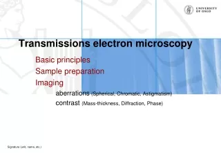
Types Transmission Electron Microscopes • Electrons travel through condenser lenses, specimen, objective lens, then projection lens before placing an optical image on a fluorescent plate • Beam speed is between 40 and 400 kiloelectron volts • Works like a projector • Specimen limited to 100 nm thickness • Cannot view surface
Types Scanning Electron Microscopes • Beam speeds between 50 and 30,000 volts • Beam interact with surface and reactions are recorded by sensors • Interacts by include producing heat, producing low energy electrons, high leveled backscattered electrons, light and/or x-ray emissions • Rotate the specimen in X,Y and Z directions
A comparison between light microscopy and two types of electron microscopy
Optical v. Electron Light Microscope Electron Microscope
Advantages • The electron microscope can be beneficial to certain studies: • Biology • Forensics • Medicine • Chemistry • Amazing resolution and magnification power (2 million times) • Chemical composition of specimen • 2D and 3D (SEM) images • Able to visualize structures that are impossible to see with other equipment • Higher depth of field
Limitations • Preservation methods must be taken, on the object such as plating, dehydration, or freezing. • Must be a small sample • Sample also must be in vacuum • Radiation • Very expensive to buy and maintain • Black and White Images